Showing posts with label Chicago. Show all posts

Wednesday, January 19, 2011

iPad May Be Best Suited For Nondiagnostic Role


Apple's iPad shows significant potential for a number of radiology applications, but primary interpretation of medical images probably isn't one of them, at least for the current generation of the tablet computer, according to researchers from the University of Maryland in Baltimore.

While its display technology is probably sufficient to handle the job, the iPad's utility for primary interpretation in radiology remains limited by visual, ergonomic, technical, and regulatory hurdles, according to Kamran Shah, MD.
"Where the potential lies in terms of existing technologies and applications [includes] [electronic medical record (EMR)] dashboards, patient interaction and consent forms, education, and as an adjunct for PACS workstations," Shah said.
Shah discussed the prospects for the iPad in radiology during a scientific session at the 2010 RSNA meeting in Chicago.
A lot to offer
At first glance, the iPad's hardware has a lot to offer. It's about one-third the weight of the average thin laptop and about three times the weight of Amazon's Kindle e-reader.
"And it's a whole lot lighter than a 40-lb PACS [display]," he said. "So it's definitely portable."
The iPad's 9.7-inch backlit display employs white LEDs in lieu of cold cathode fluorescent lamps; this provides a higher contrast ratio and increased light output compared to nonwhite LEDs, Shah said. And the LEDs are deployed in an edge-lit configuration, which facilitates a thickness of only 0.5 inches.
It also utilizes in-plane switching technology, which offers benefits such as wider viewing angles and improved color reproduction. Though it doesn't have the retina display featured on the iPhone 4, the iPad provides a resolution of 1024 x 768 pixels. That's sufficient for two frames of 512 x 512 CT or ultrasound images, Shah said.
The iPad display yields a pixel density of 132 pixels per inch, which approaches the pixel density found on 5-megapixel displays, Shah said. It also provides a resolution of 2.6 line pairs per millimeter, which meets the American College of Radiology's (ACR) display recommendation.
It has a display luminance of 300 cd/m2, which is similar to what is found on 24-inch PACS monitors.
"If you turn this up to, say, 100% max brightness and hold it in your hand, a 9.7-inch display that you're going to hold a few inches from your face is incredibly bright," he said. "This is a very bright device. Its contrast ratio is on par with what we're seeing with monitors that are being used in diagnostic applications."
Under the hood
The iPad is powered by the proprietary A4 System-on-a-Chip, which combines central processing unit (CPU), graphics processing unit (GPU), and hardware controllers. The fully autonomous GPU can render 720p video and do it in parallel with CPU tasks, Shah said.
Speed is not a high point for the iPad, however. Tests have shown that the iPad is orders of magnitude slower than a PACS workstation, Shah said.
"The iPad is anemic when compared to a PACS," he said. "This is a mobile-class processor, it's not a desktop-class processor. So it's important to recognize that."
Data connectivity is provided via 802.11n Wi-Fi, 3G capability, and Bluetooth 2.1. Memory is nonupgradeable and nonremovable, Shah said.
The iPad runs on a heavily modified version of the iPhone operating system. Multitasking was only recently added in late 2010.
"Applications will have to be rewritten to take advantage of the multitasking -- to, for example, enable DICOM viewing, speech recognition, and EMR right at the same time," he said.
As for security, the iPad offers integration with enterprise security standards and supports Microsoft Exchange ActiveSync policies. Local data are encrypted, and both local and remote data wipe is supported in case the device falls into the wrong hands, Shah said.
There are many DICOM visualization applications being developed for the iPad, all of which are going to require server-side rendering given the iPad's mobile-class processor, Shah said.
Another hurdle for diagnostic use will be the iPad's 9.7-inch display, which is going to pose ergonomic challenges if used for any protracted period of time for primary interpretation, he said.
And U.S. Food and Drug Administration (FDA) clearance for these applications remains a difficult road, Shah said.
"No DICOM visualization applications have been FDA 510(k) [cleared]," Shah said. "There have been a couple of mobile patient data applications that have been FDA 510(k) [cleared] for the iPhone and iPad, but none for image interpretation."
The iPad may also find a niche in a number of nondiagnostic image viewing applications, such as having house staff review images and reports while on rounds, Shah said.
Electronic medical record applications are another active area for the iPad, and there are more than 20 such apps in the iTunes App Store.
"The important thing about these applications is that while they do integrate with the enterprise EMR, there's the possibility of really novel integrations of data," he said. "So that you can have customized views for clinicians or for patients, [providing] a specialized dashboard for the individual user."
Clinicians could also use iPad for showing images to patients without being tethered to a PACS workstation, he said.
In addition, the iPad could be used to transform paper-based processes such as patient consent forms and questionnaires. Resident education also represents a promising application for the iPad, allowing users, for example, to interact with images while reading actual presentations, Shah said.
The iPad could also be used as an interface device, replacing other devices that are prone to failure with a multitouch device that's easier to use and less prone to fail, he said. It could be used for controlling a PACS or even for controlling an imaging modality.
"Multitouch capabilities are built into Windows 7, so it's not a huge step to imagine that this could happen," Shah said.
The iPad could be also used as inspiration for next-generation interfaces, he said.
"The whole concept of what the iPad has done for [user interface] design can be dramatic," he said.
by : Erik L Ridely

Sunday, December 19, 2010

Cause of Koulis' Death Revealed


After two days of Christ Koulis’ death, it has been confirmed that the cause of his death was a heart attack.
It was early Friday morning when the former plastic surgeon from Nashville Christ Koulis 42, died at the Northwestern Hospital in Chicago.
Chris Koulis was charged of killing his model girlfriend Lesa Buchanan in 2007. He was charged with unlawful negligence and providing her illegal drugs, whose overdose took her life.
A video tape that showed Koulis injecting Buchanan with drugs a week prior to her death cemented the charges against him and at the time of Buchanan’s death, Koulis was on
probation, but later was set free of the two year sentence court ordered him.
According to Koulis’s lawyer, David Raybin, Koulis was under stress because of the accusation he was facing following the murder of his girlfriend. This might have led to a drastic heart attack that took his life, although he was optimistic that he will win his appeal.

Thursday, December 16, 2010

Resolving Treatment Resistant Depression


About 60% of patients suffering from depression do not find any benefit from their first antidepressant. Up to 20% of patients find depression impossible to overcome even after 1 year. Assertive treatment of depression from the get go is essential to helping recover from depression.

Psychiatrists don’t have a shared definition of treatment resistant depression. However, most would agree that your depression may be treatment resistant if it has not resolved almost completely after adequate trials of at least 2 separate treatments.
You, with your doctor and/or therapist, must then consider the following strategies to beat it :

Confirm diagnosis:
Make sure that your depression is not a phase of Bipolar disorder – it can be tricky, but must be ruled out. Co-occurring addiction, anxiety or other psychiatric illness should be diagnosed and treated. Psychiatric diagnosis still remains a subjective enterprise. Get a second opinion regarding your diagnosis. Even if you like your therapist or psychiatrist, and intend to get treatment from them forever and ever, get a second opinion from a different psychiatrist.

Follow treatment recommendations:
 Take your medicine as prescribed. Maintain your frequency of psychotherapy. These things take time to work. Don’t skip doses or sessions. And don’t give up on any treatment prematurely.

Optimizing medication:
Your dose of antidepressant may need adjusting to get better results. Some antidepressants work better at higher doses. Others have a window of dosing in which they may work the best for you while having the fewest side-effects.

Switching medication:
 There are about 30 antidepressants belonging to about 8 classes available in the US. Even among patients who have had trials of multiple medicines, most have tried only 2-3 classes. Talk to your doctor about trying medicines from classes that you have not tried yet.

Adding a medication:
 If you have partial response to a given medicine, adding another medicine that is compatible with the first medicine may allow you to build on the effectiveness of the above medicine. Talk to your doctor about this. The medicine added could be another antidepressant, or a medication that is known to help antidepressants be more effective (e.g., T3, lithium).

Talk therapy (psychotherapy, counseling):
 If you are not already in psychotherapy, you must consider adding this to your treatment regimen. Psychotherapy typically does not work as quickly as medication, but tends to have lasting benefit. Ask for a referral to a therapist who treats using one of the consistently proven therapies in depression – interpersonal therapy or cognitive behavior therapy.

TMS (Transcranial Magnetic Stimulation):
 This is a treatment that involves repeatedly delivering very, very short magnetic pulses to the part of the brain that is most believed to be involved in depression. It was cleared in 2008 by the FDA as effective in those who have failed treatment with 1 adequate trial of an antidepressant. It has few side-effects, but carries with a minuscule risk of seizures. It is an outpatient treatment that does not require sedation or anesthesia.


Electroconvulsive therapy (a.k.a. electroshock therapy):
This is probably the most effective treatment for depression, but to be effective it requires that a seizure (convulsion) be triggered. Therefore, it must be conducted in a hospital setting, using sedation and muscle relaxation. It may impair memory in a lasting manner in some patients.

Most importantly, choose treatment with a physician, psychiatrist or therapist who will systematically evaluate how any chosen treatment is working. He or she should also be willing to change the medication regimen if it does not work in adequate doses over a 3-month period.
Through all of this you must also:

Practice living well:
Eat a balanced diet. Excercise some everyday (to the extent your body allows and your physician approves). Minimize smoking, drinking. Attempt to connect with loved ones frequently. None of this may be easy when you are depressed, but overcoming treatment resistance requires you to do your part to the extent that you can.


by : Dheeraj Raina
Depression Clinic of Chicago

Monday, December 13, 2010

Observed Surge in Emergency Use of CT

The use of CT in hospital emergency departments has increased at a higher rate than in other settings, which is bad news for associated radiation exposure, according to a study from Cincinnati Children's Hospital presented at the RSNA meeting.

The study was also published online today in Radiology (November 30, 2010).
David Larson, MD, and colleagues examined nationwide trends and factors associated with CT use in the emergency department using data from 1995 to 2007 National Hospital Ambulatory Medical Care Surveys. A mean of 30,044 visits were sampled for each year, and data were subcategorized according to multiple patient and hospital characteristics.
The team found that during the study time frame, the number of emergency room visits in which patients were imaged with CT increased 84% -- from 2.7 million to 16.2 million -- an average 16% increase per year. Of this growth, 3.3% was due to increased patient volume, while the remaining 12.7% was due to increased CT use, Larson said.
This overall growth represents a 5.9-fold increase and a doubling time of approximately every 4.7 years," Larson told session attendees.

The percentage of CT procedures in the emergency department grew from 2.8% in 1995 to 13.9% in 2007 -- a 4.9-fold increase, a compound annual growth rate of 14.2%, and a doubling time of 5.2 years, Larson said. CT use in the emergency department increased the most among patients 65 years of age and older, going from 6.6% in 1995 to 25.2% in 2007.
And by 2007, the end point of the study's time frame, overall use of CT had not reached an inflection point -- that is, when growth acceleration of a technology becomes negative, according to Larson.
"We did not find evidence that the increased use of CT in the emergency department was tapering," he said. "It's impossible to predict when or at what level the overall use of CT in the ED will begin to level off."
The growth partially reflects new uses for CT that were rarely used in 1995, such as side/flank imaging, abdominal imaging, and imaging for chest pain, Larson told attendees: By 2007, these indications were the most common for CT. Use of CT for side or flank pain went from 0.4% in 1995 to 43.3% in 2007; abdominal imaging went from 2% to 33%; and use of CT for chest pain increased from 1.2% to 13.7%.

This upswing in CT use means higher radiation dose in the patient population, Larson warned.
"Our study didn't directly address radiation dose, but the evidence suggests that CT-associated radiation exposure in the emergency department likely has increased even more rapidly than the number of CT examinations performed," he said.
Larson's team suggested that inappropriate CT use will decrease as more sophisticated and evidence-based decision models that address patient benefit from the exam are used at the time the decisions are made.


by : Kate Madden Yee

Walking reduces Alzheimer's progression, 1.5-tesla MRI shows


With the help of 1.5-tesla MRI, researchers at the University of Pittsburgh have found that walking 5 miles per week helps reduce the progression of mild cognitive impairment and Alzheimer's disease in adults already afflicted with the condition, according to a study presented today at the RSNA annual meeting.

The findings expand upon results of a previous study released last month, which found that walking at least 6 miles per week helped to preserve brain size and prevent the onset of dementia in healthy adults.
"Physical activity boosts the brain's resistance to Alzheimer's," said lead study author Cyrus Raji, PhD, from the university's department of radiology. "People who walked more had a slower progression of Alzheimer's disease and mild cognitive impairment over five years, and, as a result, they had a better preservation of the hippocampus, the frontal lobes, and the temporal lobes."
Patients were recruited from the Cardiovascular Health Study - Cognition Study (CHS-CS), which began approximately 20 years ago to explore coronary heart disease and stroke in people ages 65 and older.


Physical activity


Raji and colleagues analyzed the relationship between physical activity and brain structure in 426 people, including 299 healthy adults (mean age, 78 years) and 127 cognitively impaired adults (mean age, 81 years). Among the 127 cognitively impaired adults, 83 people had mild cognitive impairment and 44 adults had Alzheimer's dementia.
"Mild cognitive impairment is believed to be a very early presentation of dementia with symptoms that are not as advanced as Alzheimer's," Raji said. "As many as 50% of the people who have mild cognitive impairment go on to develop Alzheimer's."
In addition to the MRI scans, patients were given the mini-mental state exam (MMSE) to track cognitive decline over five years. Physical activity levels were correlated with MRI and MMSE results. The analysis adjusted for age, gender, body fat composition, head size, education, and other factors.
Among those who began the study as normal and eventually developed Alzheimer's and mild cognitive impairment over 10 years, the people who walked 5 miles per week -- a little less than 1 mile per day -- had similar preservation of brain volume compared to people who remained healthy longer.


image1


Alzheimer's progression


"What excites me the most is that individuals who walked 5 miles per week and developed mild cognitive impairment and Alzheimer's disease did not lose as much brain tissue over time," Raji said. "So the people who walked more had a slower progression of Alzheimer's disease and mild cognitive impairment and, as a result, they had a better preservation of the hippocampus, the frontal lobes, and the temporal lobes."
Another significant finding came from the longitudinal MMSE test results. "In the people who developed Alzheimer's disease and walked 5 miles per week, their MMSE scores went down only one point over 10 years," Raji said. "With sedentary individuals who developed Alzheimer's, their scores went down five points over 10 years."
These findings persisted even after adjusting for factors such as age, gender, race, education, and stroke. The researchers directly attributed the improvement in memory loss to the preserved hippocampal volume from walking.


image2

Raji and his colleagues plan to continue their research of what factors contribute to Alzheimer's and mild cognitive impairment by looking at diet.
"A lot of research is being done to determine if fish oil, for example, can have a beneficial effect in preventing Alzheimer's disease or reducing the risk," he said. "There have been studies done in the past suggesting that fish oil, which helps make up the building blocks of cell membranes and helps with cholesterol metabolism, might have other beneficial effects on the brain as well."


By Wayne Forrest
image 1 :The images compare the beneficial effects of physical activity on the brains of 299 healthy aging individuals to the positive relationships between exercise and brain structure in 127 people with either mild cognitive impairment or Alzheimer's. In both normal aging and cognitive impairment, physical activity preserves volume in brain areas that are important for memory, learning, and attention. These structures include the prefrontal cortex and temporal cortex. The top row of images shows these relationships in a 3D rendering of the brain, while the bottom row shows the prefrontal cortex findings in side view cutaway images of the brain.


image 2 :Side-view cutaway images show the sparing of brain volume in 299 healthy aging people (left) and 127 individuals with cognitive impairment and Alzheimer's (right). In both groups, there is more frontal lobe volume with walking about 1 mile per day, which translates to a reduced risk for cognitive impairment in the healthy aging group and slower decline in the cognitively impaired group.

Sunday, December 12, 2010

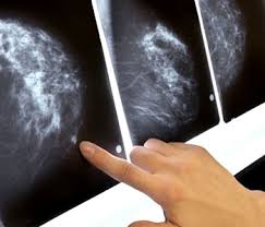
Annual mammograms reduce mastectomy risk in younger women


Annual mammograms appear to lower the risk of mastectomy in younger women, researchers reported at last week's RSNA annual meeting in Chicago.

In a study of 185 women ages 40 to 50, 22% of those who had a mammogram in the year prior to diagnosis had a mastectomy, compared with 52% who did not have a mammogram in the previous year (p = 0.02).
In addition, women who had been screened in the year prior to diagnosis had smaller tumors: 17.8 mm, on average, compared with 24 mm for those who'd had mammography more than a year ago and 28.8 mm for those who never underwent screening.
They also had less likelihood of multifocal malignancy -- 12% versus 34% for those who were never screened (p = 0.003) -- and they were less likely to have high-grade disease, at 31% versus 46%, respectively (p = 0.03).
The odds of mastectomy also correlated with the size of the tumor (p = 0.04), according to Nicholas Perry, director of the London Breast Institute at Princess Grace Hospital in London.
"Our study delivers strong clinical evidence to support annual screening of women from age 40," he said.
The researchers reviewed clinical and imaging data on women who were seen at their institute between 2003 and 2009. Only 26% had mammograms prior to their diagnosis.
Of the 48 women who had ever had a mammogram, 18 had one within the year prior to diagnosis, 15 had the exam one to two years prior, and 15 had a mammogram more than two years earlier.
Perry said that the U.K. does not recommend routine breast cancer screening for women younger than 50.
"The results are striking but should not be surprising," he said, adding that he doubted they would result in a change in recommendations.
A total of 37,000 U.S. women and 7,000 British women ages 40 to 50 are diagnosed with breast cancer each year, according to Perry. Based on these rates, yearly mammograms would spare 10,000 U.S. women and 2,000 U.K. women from mastectomy annually, he said.
When asked if the annual breast screens improved overall survival, he said longer-term studies are needed.

Wednesday, November 24, 2010

Blow


I was busted. Set up by the FBI and the DEA. That didn't bother me. Set up by Kevin Dulli and Derek Forreal to save their own asses. That didn't bother me. Sentenced to 60 years at Ottisville. That didn't bother me. I'd broken a promise. Everything I love in my life goes away.

George Jung