Showing posts with label Infectious Diseases. Show all posts

Thursday, February 6, 2025

Ebola vs. COVID-19: public health lessons from Liberia

There is a plethora of literature, addressing public health policies regarding COVID-19. The following paragraphs are excerpted from an article by Anfaara et al. published in Social Science and Medicine, January 2025

"Like elsewhere in the world, care work is gendered, with women bearing the brunt of caregiving during disease outbreaks or health emergencies. However, in Liberia, we argue that the inadequacies of the health system and past political administrations further exacerbated the care burden on women. While this aligns with our theoretical framework, it is noteworthy that despite both governments (Sirleaf and Weah) representing the elite ruling class in Liberia, our participants seem to credit one administration for being more responsive to disease containment. For instance, our participants perceived the Sirleaf administration's willingness to include grassroots organisations and other major stakeholders in Ebola management as a collective effort and a critical reason for the success of Ebola containment. This is probably because, during the initial crisis stage of Ebola, the Sirleaf administration created the National and County-level Ebola task force to brainstorm and initiate strategies to mitigate the spread of Ebola before the arrival of international support. Women's groups, in particular, were invited to be part of the national Ebola response, and there was regular Ebola messaging and communications from the government during this period; as well, on multiple occasions, Sirleaf called for an "all-hands-on-deck" approach to Ebola containment (WHO, 2015; BBC, 2014). Study participants observed that similar strategies were not fully implemented during the COVID-19 pandemic.


Certainly, the Weah administration implemented some of the post-Ebola disease management strategies to contain COVID-19. These included early surveillance, implementing a state of emergency, and restricting local and international travel (Babalola et al., 2022). Yet, participants perceived the administration's approach to COVID-19 management as unitary due to its exclusion of grassroots community-based groups in its response strategies and the government's promotion of alternative COVID-19 remedies. It is possible that the Weah's administration promotion of an alternative solution to COVID-19 containment (see News Public Trust, 2020) may have undermined vaccine uptake and reinforced conspiracy theories about the disease. This experience is not unique to Liberia; in fact, many studies have shown that leadership by positive examples reinforce scientifically proven disease management strategies while negative examples erode trust (Kutor et al., 2022; Nyenswah et al., 2016)."

Friday, January 21, 2011

A Laryngectomy Shakes This Physician To The Core


by : Itzhak Brook
As an infectious diseases physician with a special interest in head and neck infections, I had extensive experience in otolaryngological illnesses. However, when I was exposed to new, different, and challenging experiences as a neck cancer patient, I had to deal with these as a patient — not as a physician. I endured the consequences of radiation, repeated surgeries, and prolonged hospitalizations. I confronted medical errors in my care, discrimination following loss of my vocal cords, and the hardships of regaining my ability to speak.

Perhaps most importantly, I struggled to find a new meaning to my life.
Facing the diagnosis of hypo-pharyngeal carcinoma shook me to my core. I had to accept that I am not invincible, and that my life has an end. Even though the small cancer was surgically removed and I received local radiation, a recurrence two years later necessitated laryngectomy.
I encountered two types of physicians: the optimists who saw the cup half full and the pessimists who saw it half empty. I preferred those who told me the truth about the risks, potential complications, and prognosis, even when those predictions were not rosy. This is the best way one can make an educated decision about the best treatment.
Following my laryngectomy, I endured pain, weakness, the effects of narcotics, inability to eat or drink, and of course, inability to speak. I was rendered completely dependent, staying connected to an intravenous line, needing humidified, oxygenated air and constant suctioning to relieve sudden airway obstruction. All of this was an extraordinarily difficult adjustment. I understood for the first time why some patients elect to avoid heroic measures to prolong their lives, especially when their prognosis is poor.
Receiving empathetic and supportive care by the medical staff was extremely important to me. Surgeons can be impatient, rushing, and in a hurry to finish rounds, especially when they have surgeries scheduled. Most senior surgeons and many of the residents were, however, caring attentive and compassionate. I also encountered abrasive and rude physicians. On one occasion, I asked a senior resident to clean my obstructed tracheotomy tube. He reluctantly complied but did it not using a sterile technique and flushed the tube using tap water. The tube he wanted to place back was still dirty, and when I asked him to clean it better, he abrasively responded: “We call the shots here,” and left my room. I felt humiliated, helpless and angry being treated in this fashion.
Physicians need to realize how helpless and dependant their post surgical mute laryngectomees are. They require more patience and time to communicate their thoughts, questions and concerns. Their inability to speak is a very stressful and unfamiliar reality. It requires patience and support, from the medical team. Patients should be encouraged and allowed to fully express their feelings and concerns. This may be a slow and tedious process as they often need to communicate by writing. The emotional well being of the patient is a very important element of the post surgical care and ultimate recovery and adaptation to a life as a laryngectomee.
Life after laryngectomy was different and difficult, as I had to deal with new practical problems like re-learning how to eat and speak and return to a productive life. Moreover, I also confronted a difficult emotional obstacle: the constant fear that the cancer may return.
I had to continuously deal with the feeling of depression and hopelessness. Feeling depressed was surprisingly helpful when I initially faced the gloomy prospect of pending surgeries and uncertain future. It helped me to accept the potentially poor outcome. Yet I did not feel immediate depression after the surgeries because I was focused on my current situation and determined to get better. The depression, however, recurred as I had to deal with my new reality and limitations. This was enhanced by the hypothyroidism and post surgical anemia I developed.
I was determined to cope with my depression by gradually getting re-involved in professional and other activities that I had always enjoyed, including teaching, writing and even lecturing. These were instrumental in enabling me to feel better. The support of my family, a devoted social worker, responsive and caring speech and language pathologist and otolaryngologist , and members of the local support group were invaluable. However, I realized that depression can return and I will have to continuously keep it at bay.
My experiences as a patient greatly affected my approach to my patients and made me more sensitive and understanding to what they and their family are going through and feeling. I try to avoid the patterns of behaviors that were offending and distressing to me and be more compassionate and caring. I strive to model myself in the pattern of devotion, warmth and genuine care I felt from some of my caregivers. I know now better than before how critical they are. I also realize that by doing that I set an example to the physicians in training and students I teach.
Hopefully, my front-line observations will help health care providers to better understand their patients in a diligent and compassionate manner in which they were trained, and which should be the hallmark of their practice.

Dr Itzhak Brook is a Professor of Pediatrics at Georgetown University School of Medicine and author of the book My Voice: A Physician’s Personal Experience With Throat Cancer.

Tuesday, November 9, 2010

Street Life,Street Death

Here's an interesting Economist article about HIV/AIDS management in critical areas.It reveals how a derogatory attitude  hampers the fight to curb the spread of AIDS.
Pedram
At a glance, the calf-length cape that a good-looking young model swirled around on stage appeared to be made of shaggy sheepskin, wildly inappropriate for the tropical climate of Pattaya, a seaside resort in Thailand. In fact its composition was more apt, if no less uncomfortable: hundreds of dangling prophylactics. Pattaya has one of Thailand’s highest concentrations of go-go bars, massage parlours and other shop windows for the sex trade. Parts of it call to mind Sodom-on-Sea. But this “Condom Fashion Show” was not part of the sleaze. It was staged at a recent United Nations-led “consultation”. The theme—sex workers and HIV, the virus that causes AIDS—was deadly serious, and the effort to turn it into burlesque rather heroic.
The consultation differed in more fundamental ways from the usual run of international conferences. Several dozen sex workers from eight countries (Cambodia, China, Fiji, Indonesia, Myanmar, Pakistan, Papua New Guinea and Thailand) attended as full-fledged participants, not just models in the fashion show. Sex workers are on the front line in the struggle against an epidemic that in Asia still claims 350,000 new infections each year. A 2008 report prepared for the United Nations by an independent commission confirmed that “men who buy sex are the single-most powerful driving force in Asia’s HIV epidemics.” It is thought that about 10m Asian women sell sex to 75m men, who in turn have a further 50m regular partners.
A simple and effective way to cut HIV transmission is to ensure that sex workers have access to condoms, know how to use them and always do so. Yet the UN estimates that only a third of Asia’s sex workers are reached by HIV-prevention programmes. And in some countries possession of condoms is taken as evidence of involvement in prostitution, and hence a cause for harassment, extortion or detention.
Yet sex workers themselves are normally excluded—or exclude themselves—from the air-conditioned conference halls where officials and NGOs discuss their plight. One reason is a poor grasp of English, a language, one male Thai sex worker complained, that “we learned only from our clients”. More importantly, sex workers often suffer from official disdain or condescension and are seen as the problem, not the solution.
Of course, the sex industry has not been ignored in the battle against AIDS. Thailand’s early successes are perhaps best-known. It was the first country in Asia to launch a “100% condom-use programme” in the early 1990s, and it managed to cut HIV prevalence sharply. The danger now is of complacency.
The best prevention schemes involve sex workers themselves. As a Chinese delegate put it, given the chance they do a far better job of educating and helping peers than do the UN or government. Myanmar, not normally cited as an outpost of progressive policy, has an HIV-prevention scheme seen as a model. Population Services International, an international NGO, has set up a “peer-to-peer” network with 18 drop-in centres around the country. There, 350 staff, mostly former or present sex workers themselves, dispense advice, discounted condoms and treatment for sexually transmitted infections. HIV prevalence among sex workers dropped from over 30% in 2000-06 to 18% in 2008.
Still, a Burmese sex worker made a passionate intervention at the conference lamenting the stigma her profession carries. In Myanmar as elsewhere, sex workers suffer at the hands of the authorities. And the worst perpetrators are often the police and other official protectors. Take Cambodia, recognised for bringing down HIV prevalence, partly through a condom-use programme. In 2008 a law on “the Suppression of Human Trafficking and Sexual Exploitation” led to the closure of brothels and a shift of the sex trade to bars and other places, complicating measures to prevent HIV. Worse, Human Rights Watch says, it has helped the police to beat, rob and rape sex workers “with impunity”.
A group of Cambodians wrote: “Our lives and our industry have been ruined by the anti-trafficking industry. We used to work in a brothel where we have safety and solidarity… Now we are scattered on the street where it is more dangerous.”
This is an extreme example of two problems besetting efforts to help sex workers. One is that, in most places, sex work is illegal, and almost everywhere frowned upon. Indonesian delegates complained that a new “Puritanism” accompanying a slow Islamisation of public life is forcing the sex trade underground and so making it more dangerous. As our debate showed, arguments for and against decriminalising prostitution are fierce and complex. But as long as it remains a criminal act, sex workers will be vulnerable to arbitrary abuse.
Second, the debate about sex work has become drowned in a campaign against human trafficking. That campaign, however justified its goals, fosters an assumption that all sex workers ply their trade against their will. Yet most migrant sex workers have left home for good reasons of their own—among them a desire to work away from their families, and to earn more money.
Parts of the debate, such as terminology, are as old as the oldest profession. One side regards “sex worker” as a dangerously obfuscatory euphemism. The other sees “prostitute” as degrading and derogatory. One sees those who sell sex inevitably as the victims of exploitation. But those seeking to be heard in Pattaya wanted recognition as independent actors who have made their own choices. They demand to be treated with basic human dignity just as they are.

Wednesday, October 13, 2010

CR4 Amendment

Now all my attention should turn to CR4 again.Rearrangement of many sentences in discussion and finding an appealing consistency is my main task. I assume it's an enjoyable experience  . 

Monday, September 27, 2010

CR4 Wrapped Up

A great deal of pressure over this project.
Now i can reposefully focus and pursue other handy projects.

Wednesday, September 22, 2010

در مورد حادثه سوء قصد به دکتر عبدالرضا سودبخش

پلیس آگاهی تهران بزرگ در مورد حادثه سوء قصد به دکتر عبدالرضا سودبخش عضو هیئت علمی دانشگاه علوم پزشکی تهران که شب گذشته در مقابل مطب شخصی خود از طرف افراد ناشناس کشته شد اعلام کرد: تحقیقات اولیه حکایت از آن دارد که علت و انگیزه قتل شخصی بوده و وجود هرگونه انگیزه دیگر در این جنایت منتفی است.

به گزارش خبرگزاری مهر، بنا به اعلام پلیس اگاهی تهران،  در ساعت 45 : 21 مورخ 30 شهریور از طریق مرکز فوریت های پلیسی 110 وقوع یک فقره تیراندازی منجر به جرح به کلانتری 148 انقلاب اعلام شد.

با حضور مأموران کلانتری و عوامل اورژانس در بلوار کشاورز، بلافاصله فرد مصدوم به بیمارستان انتقال، اما به واسطه شدت جراحات وارده فوت می کند.

با تشکیل پرونده مقدماتی در این خصوص و به دستور قاضی کشیک دادسرای جنایی تهران، مراتب در دستور کار کارآگاهان اداره دهم ویژه قتل پلیس آگاهی تهران بزرگ قرار گرفت. با آغاز تحقیقات از سوی کارآگاهان بلافاصله هویت مقتول مورد شناسایی قرار گرفت.

تحقیقات اولیه حکایت از آن داشت که مقتول متخصص بیماری های عفونی و همچنین دانشیار دانشگاه است که در زمان خروج از مطب کار خود واقع در بلوار کشاورز مورد اصابت گلوله فرد یا افراد ناشناس قرار گرفته است.

تحقیقات اولیه حکایت از آن دارد که علت و انگیزه قتل شخصی بوده و وجود هرگونه انگیزه دیگر در این جنایت منتفی است. در حال حاضر تحقیقات تخصصی در مراحل ابتدایی قرار داشته و نتیجه اقدامات متعاقبا اعلام خواهد شد.


@tabnak.ir

Our Professor of Infectious Diseases Murdered


دكتر عبدالرضا سودبخش دانشيار بخش عفوني بيمارستان امام خميني دانشگاه علوم پزشكي تهران شب گذشته سه شنبه شب 30/6/89 ساعت 21 هنگامي كه در انتهاي كار روزانه از مطب خود خارح مي شد مورد سوءقصد فردي ناشناس قرار گرفت و به ضرب گلوله كشته شد.


از انگيزه و اهداف ضارب اطلاعي در دست نيست. مسئولين انتظامي سرگرم تحقيق پيرامون موضوعند.


دكترعبدالرضا سودبخش در سال 1328 در شهرستان بندرانزلي از توابع استان گيلان متولد شد. وي در سال 1346 موفق به اخذ مدرك ديپلم شدپ و تحصيلات عالي خود را در سال 1346 در رشته پزشكي دانشكدة پزشكي دانشگاه فردوسي مشهد آغاز كرد و در سال 1354 فارغ‌التحصيل شده بود. وي در سال 1369 نيز موفق به اخذ تخصص 

در رشتة عفوني از دانشكدة پزشكي دانشگاه علوم پزشكي تهران شده و از آن پس به عنوان عضو هيئت علمي و استاديار گروه عفوني در بخش عفوني بيمارستان امام خميني (ره) مشغول به كار شده بود وي سپس موفق يه اخذ درجه دانشياري نيز شده بود.


سمت هاي اجرائي وي شامل عضويت در كميته پژوهشي بخش عفوني، رياست درمانگاه عفوني، عضويت در كميته مورتاليته بيمارستان امام خميني (ره)، رياست كميته كنترل عفونت درمانگاهي و اورژانس مجتمع بيمارستاني امام خميني(ره) و عضويت در كميته آموزش و پژوهش فدراسيون پزشكي ورزشي ايران بود.

Friday, January 8, 2010

The Mystery Of Painful Purple Toes

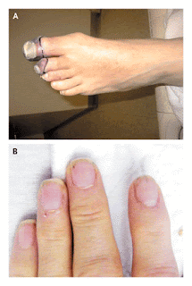
A 57-year-old man presented to the emergency department with painful purple toes. On the day of presentation, he had first been seen in another hospital but left against medical advice before the evaluation was complete. He had first noted a painful discoloration of his left great toe 2 weeks earlier. The pain and discoloration progressed to involve the left second and third toes (Figure 1A). During the 3 weeks preceding presentation, the patient also had intermittent blurry vision, intermittent chest pain, fatigue, anorexia, drenching night sweats, and a weight loss of 6.8 kg (15 lb). His roommate commented that thepatient had also been slightly confused.


Fig1A,B

This patient's painful purple toes are suggestive of peripheral arterial ischemia. The differential diagnosis includes arterial thromboembolism resulting from a hypercoagulable state or from disseminated intravascular coagulation, perhaps related to cancer; embolus of infectious or thrombotic material from endocarditis; paradoxical embolus through an intracardiac shunt; precipitation of cryoglobulins; small-vessel vasculitis; and secondary vasculitis from an underlying connective-tissue disease, such as systemic lupus erythematosus. The possibility of cholesterol emboli should also be considered, particularly in patients who have aortic aneurysms or who have recently undergone vascular procedures. Thromboangiitis obliterans, or Buerger's disease, would be a consideration if the patient smokes. His history suggests at least two and perhaps three separate vascular events in the left foot, serially affecting the digital arteries of the great, second, and third toes. Combined with the report of blurry vision and confusion, the patient's history raises the possibility that a central vascular process is affecting the peripheral and cerebral circulatory beds. The intermittent nature of the chest pain and visual symptoms suggests the possibility of sporadic emboli. Finally, the night sweats, fevers, and weight loss point to a systemic illness, such as infection, cancer, or connective-tissue disease. Specific points to review include risk factors for subacute bacterial endocarditis, such as injection-drug use, and risk factors for hypercoaguable states, such as a family history. Further history taking and physical examination should be performed to seek evidence of hypercoagulability or a systemic illness, such as endocarditis, cancer, or vasculitis.


The patient had a history of hypertension, anxiety, chronic back pain, and gastroesophageal reflux disease. While in the Marine Corps in Vietnam, he had malaria and injuries to the hip and skull. Since serving in Vietnam he had also had post-traumatic stress disorder. Two years before presentation, a screening colonoscopy detected benign polyps. His medications included alprazolam, oxycodone with acetaminophen, and esomeprazole. The patient worked for the U.S. Postal Service and was living with a friend. He reported smoking half a pack of cigarettes daily for 30 years, weekly alcohol use, and rare but ongoing intranasal cocaine use, but no intravenous drug use. He was divorced and had not recently been sexually active. There was no family history of cancer, autoimmune diseases, diabetes, or clotting disorders.
Smoking increases the risks of several cancers, including lung and pancreatic cancers, which may in turn induce a hypercoaguable state. Intranasal cocaine use can cause vasospasm, which may exacerbate digital ischemia, and is also associated with otherbehaviors that are predisposing factors for hepatitis C, a common cause of cryoglobulinemia, and acquisition of the human immunodeficiency virus (HIV). Suspicion of limb ischemia, in addition to prompting an evaluation for physical findings indicative of such systemic disease processes, calls for a thorough examination of the peripheral pulses and may warrant consultation with a vascular surgeon.
On physical examination, the patient's temperature was 36.4°C; pulse, 104 beats per minute; blood pressure, 167/86 mm Hg; respiratory rate, 14 breaths per minute; and oxygen saturation, 96% while he was breathing ambient air. No Roth's spots were seen on funduscopy. The carotid pulses were normal, without bruits, and the chest was clear on auscultation. Cardiac examination revealed a normal S1 sound and a physiologically split S2 sound, without a murmur or rub, and the presence of an S4 gallop. The abdomen was not tender, and neither the liver nor the spleen was enlarged. There was no lymphadenopathy or thrush. The dorsalis pedis and posterior tibialis pulses in the left foot were diminished but palpable, and the foot was warm. On each of the first three toes of the left foot there was a well-demarcated, cool, tender area of nonblanching, dark-purple discoloration. Capillary refill in the unaffected toes was normal. There was no rash, edema, or livedo reticularis of the legs. Distal splinter hemorrhages were noted in several fingernails (Figure 1B). There were no Janeway's lesions or Osler's nodes. Neurologic examination revealed a deficit in the left visual field. The patient received a score of 29 points out of 30 on the Mini–Mental State Examination, having difficulty only when attempting to repeat the phrase, "no ifs, ands, or buts."
The white-cell count was 15,510 per cubic millimeter, with 80% granulocytes, 14% lymphocytes, 5% monocytes, and 1% eosinophils. The hemoglobin and platelet counts were normal, and the erythrocyte sedimentation rate was 19 mm per hour. Levels of electrolytes, blood urea nitrogen, creatinine, albumin, and globulin were normal. The level of C-reactive protein was 32.1 mg per liter (reference range, 1.0 to 3.0). The serum level of creatine kinase was 40 U per liter (reference range, 41 to 266); creatine kinase MB, 2.7 ng per milliliter (reference value, <5.0);> I, 2.22 ng per milliliter (reference value, <0.04).> normalized ratio for the prothrombin time was 1.1, the partialthromboplastin time 25.7 seconds (reference range, 23.8 to 36.6), and the fibrinogen level 264 mg per deciliter (reference range, 200 to 450). A blood smear showed hypochromia; no schistocytes were identified. An electrocardiogram showed a normal sinus rhythm with ST-segment elevations in the inferior and anteroseptal leads.

The finding of a well-demarcated, discolored area on each of the three toes of the left foot supports the possibility of a separate embolus to each of the three small digital arteries feeding the affected toes, rather than a single, more proximal thrombotic or embolic event. The lesions are consistent with localized ischemia, infarction, or thromboangiitis obliterans, but the fact that the foot is warm, with palpable pulses, rules out critical limb ischemia. Although the findings on physical examination do not rule out the possibility of a small-vessel vasculitis, the absence of livedo reticularis makes the presence of cholesterol emboli unlikely. The splinter hemorrhages are consistent with systemic emboli, but these hemorrhages are a nonspecific finding that can also be associated with nail trauma, autoimmune disease, connective-tissue disease, cancer, or endocarditis. Splinter hemorrhages are more specific for subacute bacterial endocarditis when they are present in the proximal, rather than distal, nail plate.
The elevated white-cell count suggests infection, but it is a nonspecific finding. The normal coagulation studies and absence of schistocytes on the blood smear are not consistent with a diagnosis of disseminated intravascular coagulation. The combination of the elevated level of troponin I and the ST-segment abnormalities raises the possibility of an acute coronary syndrome, although the apparent involvement of two vascular territories (right and left anterior descending coronary arteries) on electrocardiography and evidence of ischemia elsewhere make emboli to the coronary circulation seem more likely. Echocardiography might identify a cardiac source of emboli and would help to gauge the extent of myocardial injury. Brain imaging is warranted to evaluate the cause of the patient's visual field deficit and slightly altered mental status.


Magnetic resonance imaging (MRI) of the brain ), including diffusion-weighted imaging, revealed multiple small lesions in the right temporal and occipital lobes, right thalamus, both parietal lobes, left frontal lobe, and both cerebellar hemispheres; there was also a large area of enhancement in the right occipital and parietal lobes. Magnetic resonance angiography of the head and neck showed decreased blood flow in the posterior communicating artery (Fig2A,B). A transthoracic echocardiogram showed a mildly thickened mitral valve, with mild regurgitation, and structurally normal aortic, tricuspid, and pulmonary valves. The left ventricular ejection fraction was normal, and therewere no abnormalities in wall motion. The results of venous ultrasonography and computed tomographic (CT) pulmonary angiography performed at the other hospital were obtained; they showed thrombi in the great saphenous veins of both legs and a small pulmonary embolus in the lower lobe of the right lung.
Fig-2A,B

The MRI findings are consistent with embolic disease of the brain, with the emboli mostly likely traveling through both the carotid and vertebrobasilar arteries from the left heart or proximal aorta. The disease process is not limited to the arterial circulation, as evidenced by the pulmonary embolus and venous thrombi of the legs. Further evaluation should focus on identifying a disease process that would explain both the venous and the arterial thrombosis, including evaluation for an underlying hypercoaguable state. Paradoxical embolism from a venous source, passing through an intracardiac shunt, could also explain the presence of both arterial and venous lesions. Although the transthoracic echocardiogram showed no evidence of a shunt, studies performed without the injection of agitated saline, such as this one, have a low sensitivity for atrial septal defects. Because of the high clinical suspicion for subacute bacterial endocarditis, further evaluation with transesophageal echocardiography is appropriate. Empiric antibiotic treatment should be initiated after blood cultures are obtained.
Emergency cardiac catheterization warrants consideration in any patient with ST-segment elevations, but in this case the normal ventricular wall motion on the echocardiogram, the presence of ST-segment abnormalities in two distinct vascular distributions, and the absence of ongoing chest pain all favor coronary emboli as the cause of the patient's symptoms and elevated troponin levels. Cerebral emboli are associated with a risk of hemorrhage, particularly if they are infectious, which in turn increases the risk of systemic anticoagulation, in addition to the anticoagulation that would be required with a percutaneous intervention performed at cardiac catheterization.
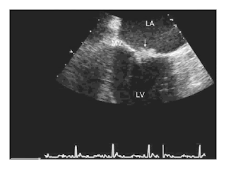
Anticoagulant therapy was deferred pending further evaluation. A transesophageal echocardiogram showed a 7-mm mobile echodensity on the atrial aspect of the posterior mitral-valve leaflet, with no evidence of perivalvular abscess or leaflet perforation, and mild-to-moderate mitral regurgitation (Figure 3). Treatment with ceftriaxone, vancomycin, and gentamicin was initiated for presumed infective endocarditis. However, multiple blood cultures — including cultures grown from samples obtained before antibiotic therapy was begun, cultures held for 2 weeks, and fungal blood cultures — were all negative. Serologic tests for HIV, coxiella, bartonella, treponema, and hepatitis B virus were negative. Serum hepatitis C virus RNA was undetectable.


Fig3- Transesophageal Echocardiogram
A midesophageal commissural view of the mitral valve shows the lesion (arrow). LA denotes left atrium, LV left ventricle, and MV mitral valve.

The negative results of tests for infection increase my suspicion that the causes of the cardiac valvular vegetations and thromboembolic disease are noninfectious. Nonbacterial thrombotic endocarditis (commonly known as marantic endocarditis) is a potential complication of connective-tissue diseases and cancer, either of which could explain the patient's weight loss and night sweats, and may also be associated with concurrent venous and arterial thromboembolism. Further studies should include assays for the lupus anticoagulant and anticardiolipin antibodies, since the antiphospholipid-antibody syndrome could explain the valvular lesion and the venous and arterial thromboses. In the absence of localizing symptoms, imaging of the abdomen and pelvis should be considered to look for evidence of cancer.
Anticardiolipin antibodies were not detected, and a test for lupus anticoagulant was negative. CT of the abdomen revealed multiple low-attenuation lesions in the liver, a finding suggestive of metastatic disease, as well as a small low-attenuation lesion in the head of the pancreas, an enlarged gastrohepatic lymph node, and bilateral wedge-shaped renal infarcts (Figure 4). CT-guided fine-needle aspiration of a liver lesion was performed, and cytologic examination of the aspirate revealed a poorly differentiated adenocarcinoma of undetermined primary origin (Figure 5). Serum levels of alpha-fetoprotein and prostate-specific antigen were normal. The level for the beta subunit of human chorionic gonadotropin was 20 mIU per milliliter (reference range, ≤1.5), and that for carcinoembryonic antigen was 4.7 ng per milliliter (reference range, ≤2.5). The serum level of the carbohydrate antigen 19-9 (CA 19-9) was 16,611 U per liter (reference value, <35).
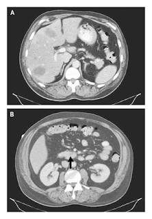


Fig4-Contrast Enhanced CT of the Abdomen
Multiple low-attenuation lesions can be seen in the liver (Panel A), and a small low-attenuation lesion is visible in the head of the pancreas (Panel B, arrow)

Figure 5


Figure 5.-Specimen from Fine-Needle Aspiration of a Liver Lesion.
Staining of the aspirate with hematoxylin and eosin shows sheets of poorly differentiated malignant cells (Panel A) and the mitotic figures and abundant clear cytoplasm that are consistent with adenocarcinoma (Panel B).


Although measurement of the CA 19-9 level should not be used as a screening test for pancreatic cancer in the general population because of its very low positive predictive value, in a patient with a pancreatic mass and suggestive clinical findings, such as this patient, a positive test result strongly supports a diagnosis of pancreatic carcinoma. Slight elevations in the levels of human chorionic gonadotropin and carcinoembryonic antigen are nonspecific and can be seen in pancreatic cancer, especially when it has metastasized to the liver. The identification of metastatic adenocarcinoma confirms the diagnosis of nonbacterial thrombotic endocarditis associated with cancer. Although concurrent arterial and venous thromboemboli are a rare complication of cancer, they are more common in cases of nonbacterial thrombotic endocarditis, as was true with this patient. Anticoagulant therapy with unfractionated heparin should be initiated to decrease the risk of recurrent thromboembolism.
Treatment with unfractionated heparin was initiated on hospital day 8; the regimen was subsequently changed to low-molecular-weight heparin. Over the course of the next week, progressive renal insufficiency, visual impairment, and episodes of psychosis with flashbacks to the Vietnam War developed. Plans for chemotherapy were deferred because of progressive multiorgan dysfunction and the poor prognosis, even with treatment. The patient requested transition to palliative care, and he died within weeks after his initial presentation. A postmortem examination was not performed.
Commentary
This patient's evaluation reveals how a careful history taking and a broad consideration of the possible causes of seemingly disparate events — including limb ischemia, blurred vision, chest pain, and weight loss — can lead to the unifying diagnosis of a systemic condition. The patient's history, the findings on physical examination, and the radiologic studies prompted consideration of a source of systemic emboli and the initiation of empirical therapy for infective endocarditis while an evaluation for noninfectious causes was performed. A key feature of this case was the presence of concurrent arterial and venous thromboemboli, which can be attributed to only a small number of unifying diagnoses.

Once a cardiac valvular mass was discovered, the clinicians chose to delay the use of systemic anticoagulation because of the risk of intracerebral hemorrhage. There is considerable controversy regarding the risk of hemorrhage when a patient has intracranial infective emboli. Some retrospective studies have shown a high risk of intracranial hemorrhage among patients with infective endocarditis and cerebral infarction who undergo anticoagulant treatment for cardiopulmonary bypass, whereas others have not. In this case, since the patient's blood cultures remained negative and radiologic imaging suggested disease that had metastasized to the liver, it became clear that nonbacterial thrombotic endocarditis was the likely diagnosis. Although the use of anticoagulation with heparin in the treatment of nonbacterial thrombotic endocarditis has not been studied in a randomized trial, it is thought to be beneficial, especially in cases that arise as a consequence of a malignant disease, and it does not seem to increase the risk of hemorrhage in association with cerebral emboli.
The prevalence of nonbacterial thrombotic endocarditis on autopsy ranges from 0.3% to 9.3%, depending on sample preparation and the prevalence of malignant disease in the source population. Although nonbacterial thrombotic endocarditis has been reported in neonates and children, frequently in association with congenital heart disease, it is most common in patients 40 years of age or older.In adults, it is often associated with cancer, but it has also been reported in association with systemic lupuserythematosus, burns, HIV infection, tuberculosis, uremia, radiation exposure, snakebites, and trauma from pulmonary catheters.When malignant disease is present, adenocarcinoma of the pancreas is cited as the most common primary cancer, as was most likely in this case; other cancers frequently found in patients with nonbacterial thrombotic endocarditis include lung, colon, and prostate cancers. The thrombophilia associated with malignant disease is thought to play an important role in the formation of valvular lesions; in a series of autopsy-proven cases of nonbacterial thrombotic endocarditis, disseminated intravascular coagulation was present in 71% of the cases. Although the laboratory findings in this patient were not typical of those associated with a consumptive coagulopathy, such as the presence of schistocytes, elevated clotting times, low fibrinogen levels, and low platelet levels, the pulmonary embolus and the venous thrombi in the legs were consistent with a hypercoaguable state induced by malignant disease.
The pathophysiology of nonbacterial thrombotic endocarditis is not well understood. Damage to the valvular endothelium is considered to be a critical first step in its pathogenesis, and patients with rheumatic or congenital heart disease are at elevated risk. Endothelial damage may be the result of high blood flow, direct trauma, immune-complex deposition, or complement activation, or it may be an elaboration of interleukin-1, interleukin-6, and tumor necrosis factor by tumor cells. The underlying thrombogenicsurface then acts as a nidus for platelet aggregation and fibrin deposition and leads to the formation of small verrucae, most of which are less than 3 mm in diameter.The valvular lesions of nonbacterial thrombotic endocarditis are usually present on the atrial surface of the mitral valve or on the ventricular surface of the aortic valve, at the point of valve coaptation. These lesions embolize frequently; the spleen, kidney, brain, and heart are the most frequently affected organs.
Current guidelines suggest that patients with nonbacterial thrombotic endocarditis and thromboembolism should be treated with full-dose heparin.As in the management of venous thromboembolism in patients with cancer, warfarin is less effective than heparin(unfractionated or low-molecular-weight) in the treatment of nonbacterial thrombotic endocarditis.Treatment of the underlying cause of the endocarditis is most likely to lead to a cure. Unfortunately, as in the present case, nonbacterial thrombotic endocarditis is often a sign of widely disseminated cancer and carries a poor prognosis.
There are no pathognomonic features in nonbacterial thrombotic endocarditis. Fever, cardiac murmur, leukocytosis, and elevated levels of C-reactive protein are present less frequently in patients with nonbacterial thrombotic endocarditis than in those with infective endocarditis. In this case, the final diagnosis of nonbacterial thrombotic endocarditis with underlying adenocarcinoma was established only after a thorough search for a cause of both the arterial and the venous thromboses.


@NEJM