Is the era of rapid growth in medical imaging procedure volume over? The volume of advanced imaging services delivered to Medicare beneficiaries decreased in 2009 -- the first decrease in 11 years, according to a study released Wednesday.
Washington, DC-based research and consulting firm the Moran Company found that the volume of advanced imaging services billed within the Medicare system decreased by 0.1% in 2009 compared with 2008, while overall imaging services declined by 7.1% for the same year-to-year comparison.
"It's pretty clear the era of very rapid growth in advanced medical imaging seems to have come to an end at this point in time," said Don Moran, president of the Moran Company. "All the data point to a leveling. It's unclear whether we will see further declines, but the prior growth of these modalities and the advantages they offer to clinicians seems to be peaking."
The study, released today by the Access to Medical Imaging Coalition (AMIC), reviewed Medicare claims data from 1999 through 2009, examining both spending and volume of advanced imaging services, such as CT, MRI, nuclear medicine, and PET, as well as overall imaging services, including mammography.
Declining Volume
The analysis also found that the total volume of mammography screenings decreased by 0.3% in 2009, compared with a 2.8% compound annual growth rate in the past decade. In addition, the total volume of dual-energy x-ray absorptiometry (DEXA) exams fell by 2.2%, while spending on this technique decreased by 16% from 2008 to 2009.
The new findings are comparable to a 2008 analysis of Medicare claims data that showed a 19.2% reduction in Medicare spending on advanced imaging from 2006 to 2007. It also revealed a significantly reduced procedure volume growth rate of only 1.9%, which was less than the overall rate of physician-payment growth.
In addition, a study presented by David C. Levin, MD, and colleagues at the 2010 RSNA meeting found that imaging procedure volume grew at a compound annual growth rate (CAGR) of 1.4% between 2005 and 2008, well down from the 4.1% CAGR experienced between 1998 and 2005.
Imaging industry observers have attributed the slowing growth rate to reimbursement cuts for medical imaging such as those enacted by the Deficit Reduction Act of 2005. The healthcare reform legislation passed in 2010 includes additional reimbursement reductions.
The study shows that "medical imaging has been decimated by these cuts," said Tim Trysla, executive director of AMIC. "The impact of these cuts, even by the government's own estimates, has 'de facto' caused access problems for patients and providers. We are very concerned about choking off the access to these lifesaving technologies."
John Patti, MD, a radiologist at Massachusetts General Hospital in Boston and chair of the American College of Radiology Board of Chancellors, said he and fellow radiologists are concerned with imaging's declining growth rate, because the Medicare population is increasing and the incidence and prevalence of disease remain the same.
Early Diagnosis
"One of the tremendous benefits of advanced imaging over the years is that it has obviated the need for more costly and evasive diagnostic evaluation," Patti said. "This reversal of trend suggests that Medicare patients may not be receiving those appropriate exams and thus the benefit of early diagnosis."
In addition, he said the decrease may have "negative downstream effects on the health of our aging citizens and on the cost of providing the more complex care that may be necessary to treat disease if it is discovered in advanced stages."
One potential victim of the decline is outpatient imaging center owners and operators. Because of reimbursement cuts for outpatient imaging, some physicians have sold or given their imaging activities to hospitals, which are reimbursed at a higher rate under the Hospital Outpatient Prospective Payment System (HOPPS). That scenario, in turn, could lead to less access to advanced imaging services.
In other study data, Medicare spending for advanced imaging services increased by less than half the spending growth for physician services overall. With a 1.2% increase in spending for advanced imaging, compared with 2.6% for services overall, imaging was one of the slowest-growing segments of the physician fee schedule in 2009.
Some Optimism
Despite the volume downturn in 2009, Trysla believes that advanced medical imaging will continue to contribute to patient care. "Medicine will not turn its back on advanced technology, especially with the benefits of early detection of disease, such as Parkinson's disease and cancer," he said. "I think you will see growth through new applications in technology, and medicine will continue to evolve from exploratory surgery toward early and more exact detection."
ACR's Patti also speculated that the dip in growth in 2009 would be temporary. "I don't think there are any physicians who are taking direct care of patients who don't understand the value of advanced imaging," he added. "And, I don't think there are any physicians who are willingly withholding [advanced imaging] from patients."
by : Wayne Forrest
Showing posts with label CT scan. Show all posts
Showing posts with label CT scan. Show all posts
Friday, February 11, 2011


Monday, December 13, 2010
Observed Surge in Emergency Use of CT
The use of CT in hospital emergency departments has increased at a higher rate than in other settings, which is bad news for associated radiation exposure, according to a study from Cincinnati Children's Hospital presented at the RSNA meeting.
The study was also published online today in Radiology (November 30, 2010).
David Larson, MD, and colleagues examined nationwide trends and factors associated with CT use in the emergency department using data from 1995 to 2007 National Hospital Ambulatory Medical Care Surveys. A mean of 30,044 visits were sampled for each year, and data were subcategorized according to multiple patient and hospital characteristics.
The team found that during the study time frame, the number of emergency room visits in which patients were imaged with CT increased 84% -- from 2.7 million to 16.2 million -- an average 16% increase per year. Of this growth, 3.3% was due to increased patient volume, while the remaining 12.7% was due to increased CT use, Larson said.
This overall growth represents a 5.9-fold increase and a doubling time of approximately every 4.7 years," Larson told session attendees.
The percentage of CT procedures in the emergency department grew from 2.8% in 1995 to 13.9% in 2007 -- a 4.9-fold increase, a compound annual growth rate of 14.2%, and a doubling time of 5.2 years, Larson said. CT use in the emergency department increased the most among patients 65 years of age and older, going from 6.6% in 1995 to 25.2% in 2007.
And by 2007, the end point of the study's time frame, overall use of CT had not reached an inflection point -- that is, when growth acceleration of a technology becomes negative, according to Larson.
"We did not find evidence that the increased use of CT in the emergency department was tapering," he said. "It's impossible to predict when or at what level the overall use of CT in the ED will begin to level off."
The growth partially reflects new uses for CT that were rarely used in 1995, such as side/flank imaging, abdominal imaging, and imaging for chest pain, Larson told attendees: By 2007, these indications were the most common for CT. Use of CT for side or flank pain went from 0.4% in 1995 to 43.3% in 2007; abdominal imaging went from 2% to 33%; and use of CT for chest pain increased from 1.2% to 13.7%.
This upswing in CT use means higher radiation dose in the patient population, Larson warned.
"Our study didn't directly address radiation dose, but the evidence suggests that CT-associated radiation exposure in the emergency department likely has increased even more rapidly than the number of CT examinations performed," he said.
Larson's team suggested that inappropriate CT use will decrease as more sophisticated and evidence-based decision models that address patient benefit from the exam are used at the time the decisions are made.
by : Kate Madden Yee
The study was also published online today in Radiology (November 30, 2010).
David Larson, MD, and colleagues examined nationwide trends and factors associated with CT use in the emergency department using data from 1995 to 2007 National Hospital Ambulatory Medical Care Surveys. A mean of 30,044 visits were sampled for each year, and data were subcategorized according to multiple patient and hospital characteristics.
The team found that during the study time frame, the number of emergency room visits in which patients were imaged with CT increased 84% -- from 2.7 million to 16.2 million -- an average 16% increase per year. Of this growth, 3.3% was due to increased patient volume, while the remaining 12.7% was due to increased CT use, Larson said.
This overall growth represents a 5.9-fold increase and a doubling time of approximately every 4.7 years," Larson told session attendees.
The percentage of CT procedures in the emergency department grew from 2.8% in 1995 to 13.9% in 2007 -- a 4.9-fold increase, a compound annual growth rate of 14.2%, and a doubling time of 5.2 years, Larson said. CT use in the emergency department increased the most among patients 65 years of age and older, going from 6.6% in 1995 to 25.2% in 2007.
And by 2007, the end point of the study's time frame, overall use of CT had not reached an inflection point -- that is, when growth acceleration of a technology becomes negative, according to Larson.
"We did not find evidence that the increased use of CT in the emergency department was tapering," he said. "It's impossible to predict when or at what level the overall use of CT in the ED will begin to level off."
The growth partially reflects new uses for CT that were rarely used in 1995, such as side/flank imaging, abdominal imaging, and imaging for chest pain, Larson told attendees: By 2007, these indications were the most common for CT. Use of CT for side or flank pain went from 0.4% in 1995 to 43.3% in 2007; abdominal imaging went from 2% to 33%; and use of CT for chest pain increased from 1.2% to 13.7%.
This upswing in CT use means higher radiation dose in the patient population, Larson warned.
"Our study didn't directly address radiation dose, but the evidence suggests that CT-associated radiation exposure in the emergency department likely has increased even more rapidly than the number of CT examinations performed," he said.
Larson's team suggested that inappropriate CT use will decrease as more sophisticated and evidence-based decision models that address patient benefit from the exam are used at the time the decisions are made.
by : Kate Madden Yee
Wednesday, December 8, 2010
Brain MRI recommended for children with hydrocephalus

In addition to not subjecting children -- especially very young children -- to radiation dose exposure, rapid MRI of the brain has been proved satisfactory for assessing ventricular size, and it does not require sedation because image acquisition can be completed in 20 seconds.
Pediatric radiologists from Children's Medical Center in Dallas made this MRI recommendation to RSNA attendees earlier this week, while delivering a sobering assessment of the cancer risk they believe their patients would face if head CT exams were continued until the children reached 20 years of age.
In a discussion of the risks of CT, it was acknowledged that the lifetime cancer risk of having a head CT scan is considerably less than with other types of CT exams, especially when low-dose protocols are utilized. Korgun Koral, MD, an associate professor of pediatric radiology at the University of Texas Southwestern Medical Center in Dallas, addressed the subject.
A 3-year-old girl has an eight in 10,000 mean lifetime risk of developing a radiation-related cancer from a head CT exam, compared to 40 in 10,000 odds with a chest CT exam. Also, the older the patient, the less the radiation dose risk. The risk of radiation-related cancer from a head CT exam is halved for a 15-year-old girl, and is halved again at age 30. (For boys, the risk is nine in 10,000, five in 10,000, and three in 10,000 at ages 3, 15, and 30, respectively.)
However, comparative assessments do not diminish the risks to a pediatric patient with shunted hydrocephalus. These patients typically have head CT exams several times a year, usually beginning at very young ages. Hydrocephalus, affects one in every 500 children, according to the National Institute of Neurological Disorders and Stroke (NINDS) in Bethesda, MD.
To treat the condition, which may be congenital or acquired, shunt systems are used to drain the cerebrospinal fluid. Treatment improvements have increased the life expectancy of these patients, Koral said.
Koral and colleagues conducted a retrospective study to estimate the lifetime attributable risk of children with shunted hydrocephalus developing a fatal cancer due to head CT for ventricular size assessment. They reviewed the medical records of Children's Medical Center from January 2009 through March 2010.
The researchers identified 150 patients, 81 of whom were female. The average age of the patients was 1.76 years, although ages ranged from 1 day to almost 19 years. To calculate the average number of exams per year that each patient underwent, the researchers counted neuroimaging studies residing in the hospital's PACS archive that were performed on these patients. In total, 211 rapid brain MRI exams and 910 head CT exams were identified. They found that 85% percent of the CT exams were performed between 6:00 a.m. and 9:00 p.m., when MRI technologists were on duty at the hospital and MRI exams could have been scheduled instead.
On average, four neuroimaging studies were performed each year, and the researchers made the assumption that these studies began for a typical patient in the second year of life. The lifetime attributable risk of developing a fatal cancer was calculated with the assumption that a patient would have four exams per year through the age of 20.
"At our hospital, the low-dose CT protocol used for hydrocephalus evaluation is 1.1 mSv, and if a standard CT protocol is used, 2.5 mSv," Koral said. "If a low dose is used consistently throughout an 18-year period, the risk of developing a fatal cancer is one in 124. If the standard dose is utilized, the risk is one in 52 patients."
"What this means is that in our current patient population, between one and three children will develop a fatal cancer if they survive to live an average adult lifetime," he said.
The researchers determined these risk estimates based on the Biologic Effects of Ionizing Radiation (BEIR) VII report, and effective doses obtained using the International Commission on Radiological Protection (ICRP) Report 103 organ weighting factors.
Whenever possible, the children's hospital uses rapid brain MRI to assess ventricular size of its patients with shunted hydrocephalus.
By Cynthia E. Keen
Friday, October 1, 2010
Schwarzenegger signs medical radiation bill into law
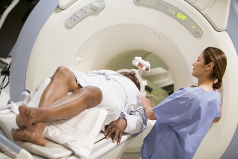
Gov. Arnold Schwarzenegger of California on Wednesday signed a medical radiation protection bill into law, paving the way for implementation of the first state law in the U.S. aimed at protecting patients from excessive radiation exposure received during CT scans and radiation therapy procedures.
SB 1237, which the governor signed without comment along with scores of other bills, will impose strict new procedures and reporting requirements to protect patients from medical radiation overdoses when it becomes effective July 1, 2012. The bill also provides an accreditation mandate for CT scanners that will take effect January 1, 2013, six months after the other provisions.
The bill requires that radiation dose be recorded on the scanned image and in a patient's health records, and that radiation overdoses be reported to patients, treating physicians, and the state Department of Public Health (DPH). It requires the same level of monitoring for therapeutic radiation used to treat cancer.
Among its reporting provisions, SB 1237 will require, for example, that medical facilities report to the DPH whenever the radiation dose for a given scan exceeds 20% of the prescribed dose, or whenever the wrong anatomic area is scanned.
"There are certainly positive steps to the bill. I think the more the public knows about the proper use of radiation, the better [it is] for the radiology community," said Robert Achermann, executive director of the Sacramento-based California Radiological Society (CRS) in an interview with AuntMinnie.com.
Many consumer and patient groups, including the California Consumer Federation, supported the bill. But CRS, which lobbies on behalf of California radiologists, has remained neutral on the legislation amid concerns over the difficulty of complying with its reporting requirements.
For this reason, Achermann called the law's delayed 2012 implementation date "critical" to implementing the best possible language.
"Delayed implementation is going to allow the radiology community, the legislators, the regulatory agents, and the [U.S. Food and Drug Administration (FDA)] to continue to explore the issues and look for the best solutions," said Achermann. CRS is continuing to work with legislators to ensure that the radiology community fully understands the bill and its requirements, and that anything that's found to be unworkable can be amended, he said.
"I'm sure this will not be a static issue in terms of what are the best ways to reduce exposure," he said. "That's what cleanup legislation is for."
Most concerning to CRS was that the Code of Federal Regulations (CFR) governing the administration of medical isotopes formed the basis of the reporting requirements in the new law, he said. Those rules are considered somewhat antiquated and not entirely appropriate to the diagnostic and therapeutic use of radiation covered by the new law, he said. From a broader perspective, it's close to impossible to do everything perfectly in a first attempt at such complex legislation as SB 1237, he said.
Between now and July 2012, the radiology community has to "ingest the bill and see the specifics," he said. "Anytime you pass new legislation, you can't have thought of everything," Achermann said. "Even though we had a lot of input from our members, there are a lot of things people don't think about. Sometimes the wording isn't appropriate and could be done better."
"This is not a California-unique issue. It's going to be used as model" for other state laws, he said.
Long road to passage
The legislation was introduced in February by State Sen. Alex Padilla (D-Pacoima) following a series of serious radiation overdoses that prompted outrage among patients and a raft of lawsuits. Last fall the California Department of Public Health learned that over an 18-month period some 260 patients at Cedars-Sinai Medical Center in Los Angeles who underwent CT perfusion scans were exposed to radiation doses eight times higher than normal.
A later review found that other California facilities, including Los Angeles County-USC Medical Center in Los Angeles; Mad River Community Hospital in Arcata; Glendale Adventist Medical Center in Glendale; Providence Saint Joseph Medical Center in Burbank; and Bakersfield Memorial Hospital in Bakersfield, among others, had also used excessive radiation doses, implicating the medical staff as well as scanner manufacturers in the errors.
The FDA is also in the process of implementing radiation protection measures that will take effect in 2012, but California's history of medical radiation accidents made the state law an urgent necessity, according to aides of Padilla.
"SB 1237 will protect patients. There is an urgent need for protocols and safeguards to prevent radiation overdoses," wrote Padilla in a September 30 statement. "This bill will provide physicians the information they need to track dosage levels, identify errors, and prevent patients from receiving overdoses of radiation."
Report : Eric Barnes
Saturday, July 31, 2010
Ewing Sarcoma
Ewing sarcoma (ES) and primitive peripheral neuroectodermal tumor (PNET) were originally described in the early 1900s as distinct clinicopathologic entities. It become evident that these entities are actually part of a spectrum of neoplastic diseases known as the Ewing sarcoma family of tumors (EFT), which also includes extraosseous ES (EES), adult neuroblastoma, malignant small-cell tumor of the thoracopulmonary region (Askin's tumor), paravertebral small-cell tumor, and atypical ES.
Because of their similar histologic and immunohistochemical characteristics and shared nonrandom chromosomal translocations, these tumors are considered to be derived from a common cell of origin. Although the histogenetic origin has been debated over the years, evidence from immunohistochemical, cytogenetic, and molecular genetic studies supports a neuroectodermal origin for all EFT
The EFT can develop in almost any bone or soft tissue, but is most common in the flat and long flat bones; patients typically present with localized pain and swelling. Although overt metastatic disease is found in fewer than 25 percent at the time of diagnosis, subclinical metastatic disease is assumed to be present in nearly all patients because of the 80 to 90 percent relapse rate in patients undergoing local therapy alone. As a result, systemic chemotherapy has evolved as an important component of treatment
Advances in multidisciplinary management of EFT over the past 30 years have resulted in a marked improvement in survival and a greater likelihood of limb-sparing surgery rather than amputation
In data derived from the Surveillance, Epidemiology and End Results (SEER) program of the National Cancer Institute, five year survival rates for patients with ES rose from 36 to 56 percent during the periods 1975 to 1984 and 1985 to 1994 .
With modern multidisciplinary treatment, long-term survival can be achieved in 70 to 80 percent of patients presenting with nonmetastatic disease
Clinical Presentation:
Primary sites — ES most often arises in the long bones of the extremities (predominantly the femur, but also the tibia, fibula and humerus), and the bones of the pelvis. The spine, hands, and feet are affected considerably less often .
Within the axial skeleton, tumors arose from the pelvis, chest wall, spine/paravertebral region, or head and neck in 45, 34, 12, and 9 percent of cases, respectively. Compared to undifferentiated ES of bone, PNET and EES more often arise within the axial rather than the appendicular skeleton
Approximately 25 percent of patients have a soft tissue primary.
Signs and Symptoms :
Patients with EFT typically present with localized pain or swelling of a few weeks or months duration
Trauma, often minor, may be the initiating event that calls attention to the lesion. The pain may be mild at first, but intensifies fairly rapidly; it may be aggravated by exercise, and is often worse at night. A distinct soft tissue mass can sometimes be appreciated. When present, it is usually firmly attached to the bone and moderately to markedly tender to palpation . Swelling of the affected limb with erythema over the mass is not uncommon.
Patients with juxta-articular lesions may present with loss of joint motion, while lesions involving the ribs can be associated with direct pleural extension and large extraosseous masses .
When the spine or sacrum are involved, nerve root irritation or compression can result in back pain, radiculopathy, or symptoms of spinal cord compression (eg, weakness or loss of bowel and/or bladder control).
Constitutional symptoms or signs, such as fever, fatigue, weight loss, or anemia, are present in about 10 to 20 percent of patients at presentation . Fever is related to cytokines produced by the tumor cells, and along with other systemic symptoms, is associated with advanced disease.
Approximately 80 percent of patients present with clinically localized disease, although as noted previously, subclinical metastatic disease is presumed to be present in nearly all. Overt metastases may become evident within weeks to months in the absence of effective therapy. The significance of this lies in the frequent delay between the onset of symptoms and diagnosis, which in one report averaged over nine months .
Patients with primary pelvic tumors are significantly more likely to present with metastatic disease compared to other sites .Other factors associated with clinically evident metastatic disease at presentation include high level of lactic dehydrogenase (LDH), the presence of fever, an interval between onset of symptoms and diagnosis less than three months, and age older than 12 years
Sites of metastatic disease at diagnosis are similar to those seen with recurrent disease; lung and bone metastases predominate, in roughly equal proportions. The spine is the most frequently involved bone.
Lung metastases represent the first site of distant spread in 70 to 80 percent of cases, and are the leading cause of death for patients with EFT.
Lymph node, liver, and brain involvement are distinctly uncommon.
Radiographic Studies :
The diagnostic work-up is usually initiated with a plain radiograph of the affected area. ES involving bone typically presents as a poorly marginated destructive lesion, most often associated with a soft tissue mass. The tumors tend to be large, and in long bones are metaphyseal or diaphyseal in location.
The radiographic appearance has been described as "permeative" or "moth-eaten", indicative of a series of finely destructive lesions that become confluent over time. The cortex at the site of the lesion is often expanded, and the periosteum displaced by the underlying tumor, resulting in the clinical sign of Codman's triangle. The characteristic periosteal reaction produces layers of reactive bone, deposited in an "onion peel" appearance.

The soft tissue component of the tumor rarely shows any calcification or ossification. Sclerosis, if present, represents a secondary bone reaction rather than the primary bone formation that characterizes osteosarcoma. A pathologic fracture is present at diagnosis in 10 to 15 percent of cases.
Compared to plain radiographs, a CT scan of the primary site better delineates the extent of cortical destruction and soft tissue disease
However, definition of tumor size, local intraosseous and extraosseous extent, and the relationship of the tumor to fascial planes, vessels, nerves, and organs is best achieved by magnetic resonance imaging (MRI)
Imaging of the entire involved bone is necessary to exclude the presence of skip lesions (ie, medullary disease within the same bone, but not in direct contiguity with the primary lesion)
Prognostic Factors :
--Disease Extent: Presence or absence of metastases,patients with bones and lungs metastases fared worse than patients with bone metastases alone
--Tumor site and size : For patients presenting with localized disease, those with axial primary tumors (ie, pelvis, rib, spine, scapula, skull, clavicle, sternum) have a worse treatment outcome than those with extremity lesions
--Response to therapy: Patients with apparently localized disease have only a 10 to 20 percent likelihood of cure if treated with surgery or radiotherapy alone; this is improved dramatically when chemotherapy is added to treatment.Both the completeness of surgical resection and the response to induction therapy are important prognostic factors. Patients who are left with significant amounts of viable tumor in the resected specimen following neoadjuvant chemotherapy do worse than those with minimal or no residual tumor.
--Histology:In most but not all studies, neither the presence of neural differentiation (eg, as in PNETs) nor extraosseous origin has a significant adverse influence on outcome
--Age: contraversy about age as prognostic factor, the five-year relapse-free survival was significantly better for children younger than 10 compared to older children
--Molecular findings:deletion of the short arm of chromosome 1p, homozygous deletions of CDKN2A and p16/p14ARF, and p53 mutations have all been associated with poor response to chemotherapy and a worse prognosis
Treatment:
Systemic chemotherapy is the mainstay of therapy ,often being used before surgery .Doxorubicin ,cyclophosphamide or ifosfamide,etoposide ,vincristine and dactinomycin are active drugs .Local treatment for the primary tumor includes surgical resection ,usually with limb salvage or radiation therapy .Patients with lesions below the elbow and below the mid-calf have a 5-year survivl rate of 80% with effective treatment .Ewing sarcoma is a curable tumor even in the presesnce of obvious metastatic disease,especially in children<11 years old.
uptodate
Harrison's Internal Medicine 2008
Because of their similar histologic and immunohistochemical characteristics and shared nonrandom chromosomal translocations, these tumors are considered to be derived from a common cell of origin. Although the histogenetic origin has been debated over the years, evidence from immunohistochemical, cytogenetic, and molecular genetic studies supports a neuroectodermal origin for all EFT
The EFT can develop in almost any bone or soft tissue, but is most common in the flat and long flat bones; patients typically present with localized pain and swelling. Although overt metastatic disease is found in fewer than 25 percent at the time of diagnosis, subclinical metastatic disease is assumed to be present in nearly all patients because of the 80 to 90 percent relapse rate in patients undergoing local therapy alone. As a result, systemic chemotherapy has evolved as an important component of treatment
Advances in multidisciplinary management of EFT over the past 30 years have resulted in a marked improvement in survival and a greater likelihood of limb-sparing surgery rather than amputation
In data derived from the Surveillance, Epidemiology and End Results (SEER) program of the National Cancer Institute, five year survival rates for patients with ES rose from 36 to 56 percent during the periods 1975 to 1984 and 1985 to 1994 .
With modern multidisciplinary treatment, long-term survival can be achieved in 70 to 80 percent of patients presenting with nonmetastatic disease
Clinical Presentation:
Primary sites — ES most often arises in the long bones of the extremities (predominantly the femur, but also the tibia, fibula and humerus), and the bones of the pelvis. The spine, hands, and feet are affected considerably less often .
Within the axial skeleton, tumors arose from the pelvis, chest wall, spine/paravertebral region, or head and neck in 45, 34, 12, and 9 percent of cases, respectively. Compared to undifferentiated ES of bone, PNET and EES more often arise within the axial rather than the appendicular skeleton
Approximately 25 percent of patients have a soft tissue primary.
Signs and Symptoms :
Patients with EFT typically present with localized pain or swelling of a few weeks or months duration
Trauma, often minor, may be the initiating event that calls attention to the lesion. The pain may be mild at first, but intensifies fairly rapidly; it may be aggravated by exercise, and is often worse at night. A distinct soft tissue mass can sometimes be appreciated. When present, it is usually firmly attached to the bone and moderately to markedly tender to palpation . Swelling of the affected limb with erythema over the mass is not uncommon.
Patients with juxta-articular lesions may present with loss of joint motion, while lesions involving the ribs can be associated with direct pleural extension and large extraosseous masses .
When the spine or sacrum are involved, nerve root irritation or compression can result in back pain, radiculopathy, or symptoms of spinal cord compression (eg, weakness or loss of bowel and/or bladder control).
Constitutional symptoms or signs, such as fever, fatigue, weight loss, or anemia, are present in about 10 to 20 percent of patients at presentation . Fever is related to cytokines produced by the tumor cells, and along with other systemic symptoms, is associated with advanced disease.
Approximately 80 percent of patients present with clinically localized disease, although as noted previously, subclinical metastatic disease is presumed to be present in nearly all. Overt metastases may become evident within weeks to months in the absence of effective therapy. The significance of this lies in the frequent delay between the onset of symptoms and diagnosis, which in one report averaged over nine months .
Patients with primary pelvic tumors are significantly more likely to present with metastatic disease compared to other sites .Other factors associated with clinically evident metastatic disease at presentation include high level of lactic dehydrogenase (LDH), the presence of fever, an interval between onset of symptoms and diagnosis less than three months, and age older than 12 years
Sites of metastatic disease at diagnosis are similar to those seen with recurrent disease; lung and bone metastases predominate, in roughly equal proportions. The spine is the most frequently involved bone.
Lung metastases represent the first site of distant spread in 70 to 80 percent of cases, and are the leading cause of death for patients with EFT.
Lymph node, liver, and brain involvement are distinctly uncommon.
Radiographic Studies :
The diagnostic work-up is usually initiated with a plain radiograph of the affected area. ES involving bone typically presents as a poorly marginated destructive lesion, most often associated with a soft tissue mass. The tumors tend to be large, and in long bones are metaphyseal or diaphyseal in location.
The radiographic appearance has been described as "permeative" or "moth-eaten", indicative of a series of finely destructive lesions that become confluent over time. The cortex at the site of the lesion is often expanded, and the periosteum displaced by the underlying tumor, resulting in the clinical sign of Codman's triangle. The characteristic periosteal reaction produces layers of reactive bone, deposited in an "onion peel" appearance.

The soft tissue component of the tumor rarely shows any calcification or ossification. Sclerosis, if present, represents a secondary bone reaction rather than the primary bone formation that characterizes osteosarcoma. A pathologic fracture is present at diagnosis in 10 to 15 percent of cases.
Compared to plain radiographs, a CT scan of the primary site better delineates the extent of cortical destruction and soft tissue disease
However, definition of tumor size, local intraosseous and extraosseous extent, and the relationship of the tumor to fascial planes, vessels, nerves, and organs is best achieved by magnetic resonance imaging (MRI)
Imaging of the entire involved bone is necessary to exclude the presence of skip lesions (ie, medullary disease within the same bone, but not in direct contiguity with the primary lesion)
Prognostic Factors :
--Disease Extent: Presence or absence of metastases,patients with bones and lungs metastases fared worse than patients with bone metastases alone
--Tumor site and size : For patients presenting with localized disease, those with axial primary tumors (ie, pelvis, rib, spine, scapula, skull, clavicle, sternum) have a worse treatment outcome than those with extremity lesions
--Response to therapy: Patients with apparently localized disease have only a 10 to 20 percent likelihood of cure if treated with surgery or radiotherapy alone; this is improved dramatically when chemotherapy is added to treatment.Both the completeness of surgical resection and the response to induction therapy are important prognostic factors. Patients who are left with significant amounts of viable tumor in the resected specimen following neoadjuvant chemotherapy do worse than those with minimal or no residual tumor.
--Histology:In most but not all studies, neither the presence of neural differentiation (eg, as in PNETs) nor extraosseous origin has a significant adverse influence on outcome
--Age: contraversy about age as prognostic factor, the five-year relapse-free survival was significantly better for children younger than 10 compared to older children
--Molecular findings:deletion of the short arm of chromosome 1p, homozygous deletions of CDKN2A and p16/p14ARF, and p53 mutations have all been associated with poor response to chemotherapy and a worse prognosis
Treatment:
Systemic chemotherapy is the mainstay of therapy ,often being used before surgery .Doxorubicin ,cyclophosphamide or ifosfamide,etoposide ,vincristine and dactinomycin are active drugs .Local treatment for the primary tumor includes surgical resection ,usually with limb salvage or radiation therapy .Patients with lesions below the elbow and below the mid-calf have a 5-year survivl rate of 80% with effective treatment .Ewing sarcoma is a curable tumor even in the presesnce of obvious metastatic disease,especially in children<11 years old.
uptodate
Harrison's Internal Medicine 2008
Friday, January 8, 2010
The Mystery Of Painful Purple Toes
A 57-year-old man presented to the emergency department with painful purple toes. On the day of presentation, he had first been seen in another hospital but left against medical advice before the evaluation was complete. He had first noted a painful discoloration of his left great toe 2 weeks earlier. The pain and discoloration progressed to involve the left second and third toes (Figure 1A). During the 3 weeks preceding presentation, the patient also had intermittent blurry vision, intermittent chest pain, fatigue, anorexia, drenching night sweats, and a weight loss of 6.8 kg (15 lb). His roommate commented that thepatient had also been slightly confused.
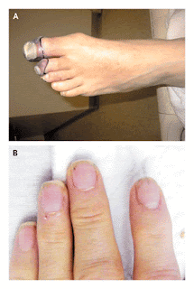
Fig1A,B
This patient's painful purple toes are suggestive of peripheral arterial ischemia. The differential diagnosis includes arterial thromboembolism resulting from a hypercoagulable state or from disseminated intravascular coagulation, perhaps related to cancer; embolus of infectious or thrombotic material from endocarditis; paradoxical embolus through an intracardiac shunt; precipitation of cryoglobulins; small-vessel vasculitis; and secondary vasculitis from an underlying connective-tissue disease, such as systemic lupus erythematosus. The possibility of cholesterol emboli should also be considered, particularly in patients who have aortic aneurysms or who have recently undergone vascular procedures. Thromboangiitis obliterans, or Buerger's disease, would be a consideration if the patient smokes. His history suggests at least two and perhaps three separate vascular events in the left foot, serially affecting the digital arteries of the great, second, and third toes. Combined with the report of blurry vision and confusion, the patient's history raises the possibility that a central vascular process is affecting the peripheral and cerebral circulatory beds. The intermittent nature of the chest pain and visual symptoms suggests the possibility of sporadic emboli. Finally, the night sweats, fevers, and weight loss point to a systemic illness, such as infection, cancer, or connective-tissue disease. Specific points to review include risk factors for subacute bacterial endocarditis, such as injection-drug use, and risk factors for hypercoaguable states, such as a family history. Further history taking and physical examination should be performed to seek evidence of hypercoagulability or a systemic illness, such as endocarditis, cancer, or vasculitis.
The patient had a history of hypertension, anxiety, chronic back pain, and gastroesophageal reflux disease. While in the Marine Corps in Vietnam, he had malaria and injuries to the hip and skull. Since serving in Vietnam he had also had post-traumatic stress disorder. Two years before presentation, a screening colonoscopy detected benign polyps. His medications included alprazolam, oxycodone with acetaminophen, and esomeprazole. The patient worked for the U.S. Postal Service and was living with a friend. He reported smoking half a pack of cigarettes daily for 30 years, weekly alcohol use, and rare but ongoing intranasal cocaine use, but no intravenous drug use. He was divorced and had not recently been sexually active. There was no family history of cancer, autoimmune diseases, diabetes, or clotting disorders.
Smoking increases the risks of several cancers, including lung and pancreatic cancers, which may in turn induce a hypercoaguable state. Intranasal cocaine use can cause vasospasm, which may exacerbate digital ischemia, and is also associated with otherbehaviors that are predisposing factors for hepatitis C, a common cause of cryoglobulinemia, and acquisition of the human immunodeficiency virus (HIV). Suspicion of limb ischemia, in addition to prompting an evaluation for physical findings indicative of such systemic disease processes, calls for a thorough examination of the peripheral pulses and may warrant consultation with a vascular surgeon.
On physical examination, the patient's temperature was 36.4°C; pulse, 104 beats per minute; blood pressure, 167/86 mm Hg; respiratory rate, 14 breaths per minute; and oxygen saturation, 96% while he was breathing ambient air. No Roth's spots were seen on funduscopy. The carotid pulses were normal, without bruits, and the chest was clear on auscultation. Cardiac examination revealed a normal S1 sound and a physiologically split S2 sound, without a murmur or rub, and the presence of an S4 gallop. The abdomen was not tender, and neither the liver nor the spleen was enlarged. There was no lymphadenopathy or thrush. The dorsalis pedis and posterior tibialis pulses in the left foot were diminished but palpable, and the foot was warm. On each of the first three toes of the left foot there was a well-demarcated, cool, tender area of nonblanching, dark-purple discoloration. Capillary refill in the unaffected toes was normal. There was no rash, edema, or livedo reticularis of the legs. Distal splinter hemorrhages were noted in several fingernails (Figure 1B). There were no Janeway's lesions or Osler's nodes. Neurologic examination revealed a deficit in the left visual field. The patient received a score of 29 points out of 30 on the Mini–Mental State Examination, having difficulty only when attempting to repeat the phrase, "no ifs, ands, or buts."
The white-cell count was 15,510 per cubic millimeter, with 80% granulocytes, 14% lymphocytes, 5% monocytes, and 1% eosinophils. The hemoglobin and platelet counts were normal, and the erythrocyte sedimentation rate was 19 mm per hour. Levels of electrolytes, blood urea nitrogen, creatinine, albumin, and globulin were normal. The level of C-reactive protein was 32.1 mg per liter (reference range, 1.0 to 3.0). The serum level of creatine kinase was 40 U per liter (reference range, 41 to 266); creatine kinase MB, 2.7 ng per milliliter (reference value, <5.0);> I, 2.22 ng per milliliter (reference value, <0.04).> normalized ratio for the prothrombin time was 1.1, the partialthromboplastin time 25.7 seconds (reference range, 23.8 to 36.6), and the fibrinogen level 264 mg per deciliter (reference range, 200 to 450). A blood smear showed hypochromia; no schistocytes were identified. An electrocardiogram showed a normal sinus rhythm with ST-segment elevations in the inferior and anteroseptal leads.
The finding of a well-demarcated, discolored area on each of the three toes of the left foot supports the possibility of a separate embolus to each of the three small digital arteries feeding the affected toes, rather than a single, more proximal thrombotic or embolic event. The lesions are consistent with localized ischemia, infarction, or thromboangiitis obliterans, but the fact that the foot is warm, with palpable pulses, rules out critical limb ischemia. Although the findings on physical examination do not rule out the possibility of a small-vessel vasculitis, the absence of livedo reticularis makes the presence of cholesterol emboli unlikely. The splinter hemorrhages are consistent with systemic emboli, but these hemorrhages are a nonspecific finding that can also be associated with nail trauma, autoimmune disease, connective-tissue disease, cancer, or endocarditis. Splinter hemorrhages are more specific for subacute bacterial endocarditis when they are present in the proximal, rather than distal, nail plate.
The elevated white-cell count suggests infection, but it is a nonspecific finding. The normal coagulation studies and absence of schistocytes on the blood smear are not consistent with a diagnosis of disseminated intravascular coagulation. The combination of the elevated level of troponin I and the ST-segment abnormalities raises the possibility of an acute coronary syndrome, although the apparent involvement of two vascular territories (right and left anterior descending coronary arteries) on electrocardiography and evidence of ischemia elsewhere make emboli to the coronary circulation seem more likely. Echocardiography might identify a cardiac source of emboli and would help to gauge the extent of myocardial injury. Brain imaging is warranted to evaluate the cause of the patient's visual field deficit and slightly altered mental status.
Magnetic resonance imaging (MRI) of the brain ), including diffusion-weighted imaging, revealed multiple small lesions in the right temporal and occipital lobes, right thalamus, both parietal lobes, left frontal lobe, and both cerebellar hemispheres; there was also a large area of enhancement in the right occipital and parietal lobes. Magnetic resonance angiography of the head and neck showed decreased blood flow in the posterior communicating artery (Fig2A,B). A transthoracic echocardiogram showed a mildly thickened mitral valve, with mild regurgitation, and structurally normal aortic, tricuspid, and pulmonary valves. The left ventricular ejection fraction was normal, and therewere no abnormalities in wall motion. The results of venous ultrasonography and computed tomographic (CT) pulmonary angiography performed at the other hospital were obtained; they showed thrombi in the great saphenous veins of both legs and a small pulmonary embolus in the lower lobe of the right lung.
 Fig-2A,B
The MRI findings are consistent with embolic disease of the brain, with the emboli mostly likely traveling through both the carotid and vertebrobasilar arteries from the left heart or proximal aorta. The disease process is not limited to the arterial circulation, as evidenced by the pulmonary embolus and venous thrombi of the legs. Further evaluation should focus on identifying a disease process that would explain both the venous and the arterial thrombosis, including evaluation for an underlying hypercoaguable state. Paradoxical embolism from a venous source, passing through an intracardiac shunt, could also explain the presence of both arterial and venous lesions. Although the transthoracic echocardiogram showed no evidence of a shunt, studies performed without the injection of agitated saline, such as this one, have a low sensitivity for atrial septal defects. Because of the high clinical suspicion for subacute bacterial endocarditis, further evaluation with transesophageal echocardiography is appropriate. Empiric antibiotic treatment should be initiated after blood cultures are obtained.
Emergency cardiac catheterization warrants consideration in any patient with ST-segment elevations, but in this case the normal ventricular wall motion on the echocardiogram, the presence of ST-segment abnormalities in two distinct vascular distributions, and the absence of ongoing chest pain all favor coronary emboli as the cause of the patient's symptoms and elevated troponin levels. Cerebral emboli are associated with a risk of hemorrhage, particularly if they are infectious, which in turn increases the risk of systemic anticoagulation, in addition to the anticoagulation that would be required with a percutaneous intervention performed at cardiac catheterization.
Anticoagulant therapy was deferred pending further evaluation. A transesophageal echocardiogram showed a 7-mm mobile echodensity on the atrial aspect of the posterior mitral-valve leaflet, with no evidence of perivalvular abscess or leaflet perforation, and mild-to-moderate mitral regurgitation (Figure 3). Treatment with ceftriaxone, vancomycin, and gentamicin was initiated for presumed infective endocarditis. However, multiple blood cultures — including cultures grown from samples obtained before antibiotic therapy was begun, cultures held for 2 weeks, and fungal blood cultures — were all negative. Serologic tests for HIV, coxiella, bartonella, treponema, and hepatitis B virus were negative. Serum hepatitis C virus RNA was undetectable.
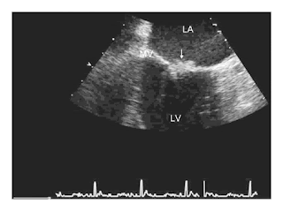 Fig3- Transesophageal Echocardiogram
A midesophageal commissural view of the mitral valve shows the lesion (arrow). LA denotes left atrium, LV left ventricle, and MV mitral valve.
The negative results of tests for infection increase my suspicion that the causes of the cardiac valvular vegetations and thromboembolic disease are noninfectious. Nonbacterial thrombotic endocarditis (commonly known as marantic endocarditis) is a potential complication of connective-tissue diseases and cancer, either of which could explain the patient's weight loss and night sweats, and may also be associated with concurrent venous and arterial thromboembolism. Further studies should include assays for the lupus anticoagulant and anticardiolipin antibodies, since the antiphospholipid-antibody syndrome could explain the valvular lesion and the venous and arterial thromboses. In the absence of localizing symptoms, imaging of the abdomen and pelvis should be considered to look for evidence of cancer.
Anticardiolipin antibodies were not detected, and a test for lupus anticoagulant was negative. CT of the abdomen revealed multiple low-attenuation lesions in the liver, a finding suggestive of metastatic disease, as well as a small low-attenuation lesion in the head of the pancreas, an enlarged gastrohepatic lymph node, and bilateral wedge-shaped renal infarcts (Figure 4). CT-guided fine-needle aspiration of a liver lesion was performed, and cytologic examination of the aspirate revealed a poorly differentiated adenocarcinoma of undetermined primary origin (Figure 5). Serum levels of alpha-fetoprotein and prostate-specific antigen were normal. The level for the beta subunit of human chorionic gonadotropin was 20 mIU per milliliter (reference range,
 1.5), and that for carcinoembryonic antigen was 4.7 ng per milliliter (reference range, 2.5). The serum level of the carbohydrate antigen 19-9 (CA 19-9) was 16,611 U per liter (reference value, <35).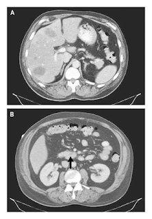
Fig4-Contrast Enhanced CT of the Abdomen
Multiple low-attenuation lesions can be seen in the liver (Panel A), and a small low-attenuation lesion is visible in the head of the pancreas (Panel B, arrow)

Figure 5.-Specimen from Fine-Needle Aspiration of a Liver Lesion.
Staining of the aspirate with hematoxylin and eosin shows sheets of poorly differentiated malignant cells (Panel A) and the mitotic figures and abundant clear cytoplasm that are consistent with adenocarcinoma (Panel B).
Although measurement of the CA 19-9 level should not be used as a screening test for pancreatic cancer in the general population because of its very low positive predictive value, in a patient with a pancreatic mass and suggestive clinical findings, such as this patient, a positive test result strongly supports a diagnosis of pancreatic carcinoma. Slight elevations in the levels of human chorionic gonadotropin and carcinoembryonic antigen are nonspecific and can be seen in pancreatic cancer, especially when it has metastasized to the liver. The identification of metastatic adenocarcinoma confirms the diagnosis of nonbacterial thrombotic endocarditis associated with cancer. Although concurrent arterial and venous thromboemboli are a rare complication of cancer, they are more common in cases of nonbacterial thrombotic endocarditis, as was true with this patient. Anticoagulant therapy with unfractionated heparin should be initiated to decrease the risk of recurrent thromboembolism.
Treatment with unfractionated heparin was initiated on hospital day 8; the regimen was subsequently changed to low-molecular-weight heparin. Over the course of the next week, progressive renal insufficiency, visual impairment, and episodes of psychosis with flashbacks to the Vietnam War developed. Plans for chemotherapy were deferred because of progressive multiorgan dysfunction and the poor prognosis, even with treatment. The patient requested transition to palliative care, and he died within weeks after his initial presentation. A postmortem examination was not performed.
Commentary
This patient's evaluation reveals how a careful history taking and a broad consideration of the possible causes of seemingly disparate events — including limb ischemia, blurred vision, chest pain, and weight loss — can lead to the unifying diagnosis of a systemic condition. The patient's history, the findings on physical examination, and the radiologic studies prompted consideration of a source of systemic emboli and the initiation of empirical therapy for infective endocarditis while an evaluation for noninfectious causes was performed. A key feature of this case was the presence of concurrent arterial and venous thromboemboli, which can be attributed to only a small number of unifying diagnoses.
Once a cardiac valvular mass was discovered, the clinicians chose to delay the use of systemic anticoagulation because of the risk of intracerebral hemorrhage. There is considerable controversy regarding the risk of hemorrhage when a patient has intracranial infective emboli. Some retrospective studies have shown a high risk of intracranial hemorrhage among patients with infective endocarditis and cerebral infarction who undergo anticoagulant treatment for cardiopulmonary bypass, whereas others have not. In this case, since the patient's blood cultures remained negative and radiologic imaging suggested disease that had metastasized to the liver, it became clear that nonbacterial thrombotic endocarditis was the likely diagnosis. Although the use of anticoagulation with heparin in the treatment of nonbacterial thrombotic endocarditis has not been studied in a randomized trial, it is thought to be beneficial, especially in cases that arise as a consequence of a malignant disease, and it does not seem to increase the risk of hemorrhage in association with cerebral emboli.
The prevalence of nonbacterial thrombotic endocarditis on autopsy ranges from 0.3% to 9.3%, depending on sample preparation and the prevalence of malignant disease in the source population. Although nonbacterial thrombotic endocarditis has been reported in neonates and children, frequently in association with congenital heart disease, it is most common in patients 40 years of age or older.In adults, it is often associated with cancer, but it has also been reported in association with systemic lupuserythematosus, burns, HIV infection, tuberculosis, uremia, radiation exposure, snakebites, and trauma from pulmonary catheters.When malignant disease is present, adenocarcinoma of the pancreas is cited as the most common primary cancer, as was most likely in this case; other cancers frequently found in patients with nonbacterial thrombotic endocarditis include lung, colon, and prostate cancers. The thrombophilia associated with malignant disease is thought to play an important role in the formation of valvular lesions; in a series of autopsy-proven cases of nonbacterial thrombotic endocarditis, disseminated intravascular coagulation was present in 71% of the cases. Although the laboratory findings in this patient were not typical of those associated with a consumptive coagulopathy, such as the presence of schistocytes, elevated clotting times, low fibrinogen levels, and low platelet levels, the pulmonary embolus and the venous thrombi in the legs were consistent with a hypercoaguable state induced by malignant disease.
The pathophysiology of nonbacterial thrombotic endocarditis is not well understood. Damage to the valvular endothelium is considered to be a critical first step in its pathogenesis, and patients with rheumatic or congenital heart disease are at elevated risk. Endothelial damage may be the result of high blood flow, direct trauma, immune-complex deposition, or complement activation, or it may be an elaboration of interleukin-1, interleukin-6, and tumor necrosis factor by tumor cells. The underlying thrombogenicsurface then acts as a nidus for platelet aggregation and fibrin deposition and leads to the formation of small verrucae, most of which are less than 3 mm in diameter.The valvular lesions of nonbacterial thrombotic endocarditis are usually present on the atrial surface of the mitral valve or on the ventricular surface of the aortic valve, at the point of valve coaptation. These lesions embolize frequently; the spleen, kidney, brain, and heart are the most frequently affected organs.
Current guidelines suggest that patients with nonbacterial thrombotic endocarditis and thromboembolism should be treated with full-dose heparin.As in the management of venous thromboembolism in patients with cancer, warfarin is less effective than heparin(unfractionated or low-molecular-weight) in the treatment of nonbacterial thrombotic endocarditis.Treatment of the underlying cause of the endocarditis is most likely to lead to a cure. Unfortunately, as in the present case, nonbacterial thrombotic endocarditis is often a sign of widely disseminated cancer and carries a poor prognosis.
There are no pathognomonic features in nonbacterial thrombotic endocarditis. Fever, cardiac murmur, leukocytosis, and elevated levels of C-reactive protein are present less frequently in patients with nonbacterial thrombotic endocarditis than in those with infective endocarditis. In this case, the final diagnosis of nonbacterial thrombotic endocarditis with underlying adenocarcinoma was established only after a thorough search for a cause of both the arterial and the venous thromboses.
@NEJM
Subscribe to:
Posts (Atom)