Showing posts with label Mamography. Show all posts

Monday, May 16, 2011

Phyllodes Tumor


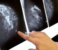
Findings: Mammogram shows a lobular hyper dense mass with partially circumscribed and partially obscured margins. No calcifications visible

Meaning : leaf-like in Greek, phyllodes tumors demonstrate papillary growth of epithelial lined stroma
Key point: Large rapidly growing circumscribed mass without calcifications
Mammography :Phyllodes tumors appear as a dense, round or oval masses with circumscribed or lobulated borders on mammography. Indistinct margins favor malignant transformation. Calcifications are rare but when present are coarse.
Ultrasonography: demonstrates an oval, round, or lobulated hypoechoic mass. Cystic spaces favor malignancy. Increased vascularity can be common .

T1W1/T2W1 :Phyllodes tumors appear heterogeneously hypointense on T1WI with slit like areas of increased T2WI representing fluid. Enhancement is typically rapid and suspicious kinetics can be observed in approximately 33% of cases.
Differential Diagnosis:
Typically occurring in younger women, fibroadenomas also appear as oval or lobulated mass but have dense, coarse calcifications, homogeneous echogenicity, and more moderate enhancement characteristics.
While there is some overlap with phyllodes tumors demonstrating malignant transformation, breast carcinoma is more likely to demonstrate indistinct margins. Pleomorphic calcifications also favor carcinoma.
Primary sarcoma of the breast is distinguished from phyllodes tumors by the absence of epithelial components. The clinical course of primary sarcoma of the breast is similar to malignant phyllodes tumors.


Ultrasound shows an irregularly shaped mass with heterogeneous echogenicity and ill-defined borders.
Treatment: is by surgical excision with greater than 1 cm margins. Mastectomy may be required for large tumors. Axillary node dissection is usually unnecessary. With respect to adjuvant therapy, radiation reduces local recurrence. Chemotherapy has demonstrated no benefit.

Friday, February 11, 2011

Imaging's Torrid Growth Rate Is Slowing

 Is the era of rapid growth in medical imaging procedure volume over? The volume of advanced imaging services delivered to Medicare beneficiaries decreased in 2009 -- the first decrease in 11 years, according to a study released Wednesday.
Washington, DC-based research and consulting firm the Moran Company found that the volume of advanced imaging services billed within the Medicare system decreased by 0.1% in 2009 compared with 2008, while overall imaging services declined by 7.1% for the same year-to-year comparison.

"It's pretty clear the era of very rapid growth in advanced medical imaging seems to have come to an end at this point in time," said Don Moran, president of the Moran Company. "All the data point to a leveling. It's unclear whether we will see further declines, but the prior growth of these modalities and the advantages they offer to clinicians seems to be peaking."
The study, released today by the Access to Medical Imaging Coalition (AMIC), reviewed Medicare claims data from 1999 through 2009, examining both spending and volume of advanced imaging services, such as CT, MRI, nuclear medicine, and PET, as well as overall imaging services, including mammography.

Declining Volume

The analysis also found that the total volume of mammography screenings decreased by 0.3% in 2009, compared with a 2.8% compound annual growth rate in the past decade. In addition, the total volume of dual-energy x-ray absorptiometry (DEXA) exams fell by 2.2%, while spending on this technique decreased by 16% from 2008 to 2009.
The new findings are comparable to a 2008 analysis of Medicare claims data that showed a 19.2% reduction in Medicare spending on advanced imaging from 2006 to 2007. It also revealed a significantly reduced procedure volume growth rate of only 1.9%, which was less than the overall rate of physician-payment growth.
In addition, a study presented by David C. Levin, MD, and colleagues at the 2010 RSNA meeting found that imaging procedure volume grew at a compound annual growth rate (CAGR) of 1.4% between 2005 and 2008, well down from the 4.1% CAGR experienced between 1998 and 2005.
Imaging industry observers have attributed the slowing growth rate to reimbursement cuts for medical imaging such as those enacted by the Deficit Reduction Act of 2005. The healthcare reform legislation passed in 2010 includes additional reimbursement reductions.
The study shows that "medical imaging has been decimated by these cuts," said Tim Trysla, executive director of AMIC. "The impact of these cuts, even by the government's own estimates, has 'de facto' caused access problems for patients and providers. We are very concerned about choking off the access to these lifesaving technologies."
John Patti, MD, a radiologist at Massachusetts General Hospital in Boston and chair of the American College of Radiology Board of Chancellors, said he and fellow radiologists are concerned with imaging's declining growth rate, because the Medicare population is increasing and the incidence and prevalence of disease remain the same.

Early Diagnosis

"One of the tremendous benefits of advanced imaging over the years is that it has obviated the need for more costly and evasive diagnostic evaluation," Patti said. "This reversal of trend suggests that Medicare patients may not be receiving those appropriate exams and thus the benefit of early diagnosis."
In addition, he said the decrease may have "negative downstream effects on the health of our aging citizens and on the cost of providing the more complex care that may be necessary to treat disease if it is discovered in advanced stages."
One potential victim of the decline is outpatient imaging center owners and operators. Because of reimbursement cuts for outpatient imaging, some physicians have sold or given their imaging activities to hospitals, which are reimbursed at a higher rate under the Hospital Outpatient Prospective Payment System (HOPPS). That scenario, in turn, could lead to less access to advanced imaging services.
In other study data, Medicare spending for advanced imaging services increased by less than half the spending growth for physician services overall. With a 1.2% increase in spending for advanced imaging, compared with 2.6% for services overall, imaging was one of the slowest-growing segments of the physician fee schedule in 2009.


Some Optimism
Despite the volume downturn in 2009, Trysla believes that advanced medical imaging will continue to contribute to patient care. "Medicine will not turn its back on advanced technology, especially with the benefits of early detection of disease, such as Parkinson's disease and cancer," he said. "I think you will see growth through new applications in technology, and medicine will continue to evolve from exploratory surgery toward early and more exact detection."
ACR's Patti also speculated that the dip in growth in 2009 would be temporary. "I don't think there are any physicians who are taking direct care of patients who don't understand the value of advanced imaging," he added. "And, I don't think there are any physicians who are willingly withholding [advanced imaging] from patients."


by : Wayne Forrest

Saturday, January 15, 2011

On Breast Elastography


Breast ultrasound elastography is 12% more sensitive than MR diffusion-weighted imaging (DWI) in determining malignancy of breast masses assessed as BI-RADS category 4, and it's almost 10% more accurate, according to a new study published in the January American Journal of Roentgenology.
Clinicians currently use B-mode sonography and dynamic contrast-enhanced MRI to classify breast lesions based on the standard BI-RADS categorizations. But newer techniques such as ultrasound elastography -- which assesses the softness or stiffness of breast tissue -- and DWI-MRI are being evaluated as adjuncts to these modalities in the hope of better identifying the character of a breast lesion.
Hiroko Satake, MD, of Nagoya University School of Medicine in Japan, and colleagues compared the abilities of ultrasound elastography and DWI-MRI to predict malignancy of breast masses. They found that not only was elastography more sensitive overall, it was more sensitive with lesions smaller than 1 cm -- more than 17% compared to DWI-MRI. In addition, it was more than 10% more accurate (AJR, January 2011, Vol. 196:1, pp. 202-209).
"Because malignant tumors predominantly are harder than benign tissues, [ultrasound elastography] significantly improves the differentiation between benign and malignant tissue," Satake and colleagues wrote. "[Our results] suggest that ultrasound elastography could be used to prevent unnecessary biopsies."
Satake's group included 115 breast masses categorized as BI-RADS 4 or 5; the masses were assessed according to combined findings from mammography, B-mode sonography, and dynamic contrast-enhanced MRI. Two radiologists retrospectively evaluated the elasticity scores of the masses using ultrasound elastography and the apparent diffusion coefficient (ADC) values using DWI-MRI.
"By accurately identifying benign tumors with imaging, we may be able to avoid sending patients for unnecessary biopsies," Satake and colleagues wrote. "Based on the results of our study, we recommend that patients with BI-RADS 4 masses should undergo biopsy if their ultrasound elasticity score is 4 or 5."
Of the 115 breast masses included in the study, 88 were malignant and 27 were benign. The mean diameter of the malignant lesions was 16.1 mm, the team found. The researchers compared BI-RADS assessment categories, elasticity scores, and ADC values between the benign and malignant masses. A lesion's elasticity score proved to be more sensitive and accurate in predicting malignancy than its ADC value, both overall and with lesions smaller than 1 cm.


by : Kate Madden Yee

Sunday, December 12, 2010

Annual mammograms reduce mastectomy risk in younger women


Annual mammograms appear to lower the risk of mastectomy in younger women, researchers reported at last week's RSNA annual meeting in Chicago.

In a study of 185 women ages 40 to 50, 22% of those who had a mammogram in the year prior to diagnosis had a mastectomy, compared with 52% who did not have a mammogram in the previous year (p = 0.02).
In addition, women who had been screened in the year prior to diagnosis had smaller tumors: 17.8 mm, on average, compared with 24 mm for those who'd had mammography more than a year ago and 28.8 mm for those who never underwent screening.
They also had less likelihood of multifocal malignancy -- 12% versus 34% for those who were never screened (p = 0.003) -- and they were less likely to have high-grade disease, at 31% versus 46%, respectively (p = 0.03).
The odds of mastectomy also correlated with the size of the tumor (p = 0.04), according to Nicholas Perry, director of the London Breast Institute at Princess Grace Hospital in London.
"Our study delivers strong clinical evidence to support annual screening of women from age 40," he said.
The researchers reviewed clinical and imaging data on women who were seen at their institute between 2003 and 2009. Only 26% had mammograms prior to their diagnosis.
Of the 48 women who had ever had a mammogram, 18 had one within the year prior to diagnosis, 15 had the exam one to two years prior, and 15 had a mammogram more than two years earlier.
Perry said that the U.K. does not recommend routine breast cancer screening for women younger than 50.
"The results are striking but should not be surprising," he said, adding that he doubted they would result in a change in recommendations.
A total of 37,000 U.S. women and 7,000 British women ages 40 to 50 are diagnosed with breast cancer each year, according to Perry. Based on these rates, yearly mammograms would spare 10,000 U.S. women and 2,000 U.K. women from mastectomy annually, he said.
When asked if the annual breast screens improved overall survival, he said longer-term studies are needed.