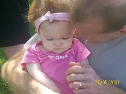
by : Danielle Leach, MPA
“A true friend walks in when everyone else walks out.”
I read that on a magnet on my friend’s refrigerator recently and the simple power of that saying brought me to tears. I have learned that lesson of true friends since my son’s diagnosis of cancer in 2007.
Anyone who has faced a serious illness as a patient or a caregiver knows that you quickly learn who your friends are. They are the ones who are there, who listen instead of trying to fix things, who are present for you in any way you need them. Some people you love will disappoint and not rise to the occasion, and some people you never expected will be your biggest supporters.
It is hard not to resent people who are there in the crisis, and then leave once the immediate crisis is over. There are people who are not there for the long haul, for the good and the bad that a disease may bring. The initial drama draws everyone in, but sends them running afterward.
I have learned, especially when you are living a nightmare, that it takes a special person to stay with you throughout the crisis. A person who keeps checking in and knows the journey is not necessarily over once you are in remission, or when your loved one has passed away. When my son Mason had brain cancer, our family found our true friends. We were surprised by many who walked out, but also by how many true friends walked into our lives because of Mason’s illness. We have learned even after Mason’s death, even three years later, we continue to go through this process of discovering our true friends.
Some people are not capable of handling personal difficulties. We, as patients and caregivers, need to understand not everyone has the capacity or tools to handle a crisis of another. This knowledge does not make it any easier for us as we wade through process of dealing with disease. As a director at Inspire, a company that creates and manages online patient support communities, I see regularly the comments of patients and caregivers who talk about friendships won and lost since diagnosis. Some are surprised and profoundly saddened by the lack of support from those expected to help the most. However, many happily note those friends, family, and even strangers who surprise them with support in a time of great need.
I recall reading about a Florida woman, whose teenage son was undergoing chemo, wrote that her friends avoided her upon learning about her son’s cancer diagnosis. “It’s almost like they were afraid they could catch it,” she said.
Another, a bladder cancer survivor from New Jersey, observed, “A lot of people walk out. . . a good 50% of my ‘pre-cancer’ friends I have never heard from again.” He went on to say, “In my case, I am lucky. I have all strong ones, having cut weak relations a long time ago. I keep only the cream of the crop.”
Sometimes finding others who are dealing with the same issues can be the most helpful strategy. You can often talk online more frankly and honestly with them than with some loved ones or friends. Dealing with an illness can be a lonely and scary process. Participating in support communities often help alleviate some of that loneliness. I have seen repeatedly how these connections are a powerful tool and establish strong personal friendships among members.
If you’re a patient or caregiver, look for the people who are true friends and hold those people close. Craft a strong support network–both in person and online. If you have a chance to do so, be the kind of true friend people are often searching for in their lives when they need it the most.
Danielle Leach is Director of Partnerships at Inspire and is founder of the Mason Leach Superstar Fund, in memory of her son, Mason, who died of pediatric medulloblastoma in 2007.
by : Danielle Leach, MPA
“A true friend walks in when everyone else walks out.”
I read that on a magnet on my friend’s refrigerator recently and the simple power of that saying brought me to tears. I have learned that lesson of true friends since my son’s diagnosis of cancer in 2007.
Anyone who has faced a serious illness as a patient or a caregiver knows that you quickly learn who your friends are. They are the ones who are there, who listen instead of trying to fix things, who are present for you in any way you need them. Some people you love will disappoint and not rise to the occasion, and some people you never expected will be your biggest supporters.
It is hard not to resent people who are there in the crisis, and then leave once the immediate crisis is over. There are people who are not there for the long haul, for the good and the bad that a disease may bring. The initial drama draws everyone in, but sends them running afterward.
I have learned, especially when you are living a nightmare, that it takes a special person to stay with you throughout the crisis. A person who keeps checking in and knows the journey is not necessarily over once you are in remission, or when your loved one has passed away. When my son Mason had brain cancer, our family found our true friends. We were surprised by many who walked out, but also by how many true friends walked into our lives because of Mason’s illness. We have learned even after Mason’s death, even three years later, we continue to go through this process of discovering our true friends.
Some people are not capable of handling personal difficulties. We, as patients and caregivers, need to understand not everyone has the capacity or tools to handle a crisis of another. This knowledge does not make it any easier for us as we wade through process of dealing with disease. As a director at Inspire, a company that creates and manages online patient support communities, I see regularly the comments of patients and caregivers who talk about friendships won and lost since diagnosis. Some are surprised and profoundly saddened by the lack of support from those expected to help the most. However, many happily note those friends, family, and even strangers who surprise them with support in a time of great need.
I recall reading about a Florida woman, whose teenage son was undergoing chemo, wrote that her friends avoided her upon learning about her son’s cancer diagnosis. “It’s almost like they were afraid they could catch it,” she said.
Another, a bladder cancer survivor from New Jersey, observed, “A lot of people walk out. . . a good 50% of my ‘pre-cancer’ friends I have never heard from again.” He went on to say, “In my case, I am lucky. I have all strong ones, having cut weak relations a long time ago. I keep only the cream of the crop.”
Sometimes finding others who are dealing with the same issues can be the most helpful strategy. You can often talk online more frankly and honestly with them than with some loved ones or friends. Dealing with an illness can be a lonely and scary process. Participating in support communities often help alleviate some of that loneliness. I have seen repeatedly how these connections are a powerful tool and establish strong personal friendships among members.
If you’re a patient or caregiver, look for the people who are true friends and hold those people close. Craft a strong support network–both in person and online. If you have a chance to do so, be the kind of true friend people are often searching for in their lives when they need it the most.
Danielle Leach is Director of Partnerships at Inspire and is founder of the Mason Leach Superstar Fund, in memory of her son, Mason, who died of pediatric medulloblastoma in 2007.
If you think about it, the comparison of doctors to mechanics is pretty apt. Given the specialized nature of their work and the general helplessness of most patients, doctors, like mechanics, are rarely held accountable for their actions. Sure, for the most part, doctors have your best interest at heart, but like in any profession, there are both good and bad apples. According to the National Patient Safety Foundation, 42 percent of people believed they had personally experienced a medical mistake. Additionally, numerous statistics have shown that a staggering amount of people die each year due to medical error. The following doctors made such blatant mistakes and/or lapses in judgment that they, at the very least, have a reputation that garnered them recognition on this list.
1.Dr. Earl Bradley, Monster: Nothing is worse than violating the trust of a child, which is why Bradley, a former pediatrician from Lewes, Delaware, is such a monster. In February of 2010, he was indicted on 471 charges of child sex abuse involving 103 children. Initial allegations against Bradley surfaced during the mid-’90s, but the hospital for which he worked was unable to verify the claims. They were enough, however, to prompt him to move to Delaware, where more allegations surfaced a decade later, including that he had abused his own son. Bradley was initially arrested in December of 2009 after a year-long investigation, which prompted the indictments two months later.
2.Dr. Robert Ricketson, Screw Up: Ricketson is no longer practicing medicine and the world is better for it. During a spine operation he performed on 73-year-old Arturo Iturralde in 2003, Ricketson intentionally inserted a piece of a screwdriver into the patient’s spine instead of a titanium rod. The rod he had intended to use went missing prior to the surgery, so he took it upon himself to improvise. As a result, Iturralde endured three additional surgeries to insert the proper rod and correct complications. After one of the operations, pieces of the screwdriver were recovered by nurses and the family was alerted. Iturralde became a paraplegic and died two years later, and his family was awarded $5.6 million in a malpractice lawsuit in 2006. During the aftermath, it was discovered that Ricketson’s medical license had been suspended in Oklahoma and Texas and he was denied consideration for a medical license in Kansas in 2002, a year after the botched surgery. He had previously been sued for malpractice several times and had history of narcotics abuse. Not a very impressive resume to say the least.
3.Dr. Jan Adams, Plastic Surgeon Imposter: Adams is most famous for performing the breast augmentation surgery on Donda West, Kanye West’s mom, that resulted in her death in 2007. But that’s not the only blemish on his record. In a malpractice suit against Adams, a previous patient claimed that she didn’t receive proper preoperative or postoperative care, leading to an infection that needed two more surgeries. What’s more, one patient claimed he got her drunk after a surgery and impregnated her, and another claimed a surgical sponge was left inside of her after a breast augmentation. Adams, who attended Harvard University, never received his diploma despite apparently completing his academic requirements. He’s also not a board certified plastic surgeon despite claiming to be one. How was this guy getting work?
4.Julie Ponder and Connell Watkins, Child Killers: Alternative medicine can be a risky proposition given the unproven nature of many of its methods. Attachment therapy in particular can yield tragic results when taken to an extreme. The treatment is used to remedy attachment disorders primarily suffered by adopted and fostered children who persistently misbehave or display little affection toward their new caregivers. In 2000, 10-year-old Candace Newmaker was killed during such treatment when she was suffocated during an intensive rebirthing session. Wrapped in a flannel sheet by psychotherapists Julie Ponder and Connell Watkins, she told to free herself from it while Ponder and Watkins held her down. She pleaded for her life, but was told by Ponder "You want to die? OK, then die. Go ahead, die right now." Newmaker was declared brain dead the next day due to asphyxia. The entire session was videotaped and presented as evidence against Ponder and Watkins, who were each given 16-year prison sentences — Watkins was paroled in 2008 after serving seven years.
5.Dr. James Burt, Ghastly Gyno: The notorious Dr. Burt was exposed for his harmful and downright bizarre practices in the late 1980s when numerous former patients came forward and initiated lawsuits. Beginning in the late 1960s, Burt took it upon himself to perform "love surgeries" in which he altered his patients’ vulvas without their consent. He justified his work in a book he authored in 1975, explaining that "Women are structurally inadequate for intercourse. This is a pathological condition amenable by surgery." He stated the procedure turns them into "horny little mice," though in reality, many of them suffered sexual dysfunction, infection and required corrective surgeries as a result. A $21 million suit was filed against Burt that couldn’t have come close to covering the physical and emotional damages endured by the at least 40 women he hurt.
6.Dr. Cecil Jacobson, Seed Spreader: As a fertility doctor, it was Dr. Jacobson’s duty to assist women in conceiving, and in the 1980s, it appeared he was doing a pretty good job. His patients reported high success rates due to his use of hCG, a hormone released during pregnancy that causes the typical bodily changes. During the supposed pregnancies initiated by hCG, he would identify the fetuses during an ultrasound, but they would usually "die" after about three months. Patients who suspected something was amiss informed a local television station, which investigated and exposed Jacobson. In the process, they discovered he used his semen to artificially inseminate patients who were told they were matched with an anonymous donor. Genetic testing later showed that he was the biological father of at least seven of his patients’ children. Jacobson, who won the Ig Nobel Prize for Biology in 1992 and claimed to have successfully oversaw the impregnation of a male baboon in the 1960s, was stripped of medical license and sentenced to five years in prison.
7.Dr. Rolando R. Sanchez, Accidental Amputator: To be fair to Dr. Sanchez, he has done a fine job of rehabilitating his reputation after the costly mistake he oversaw in 1995 — in fact, he’s still practicing. Even still, it’s the kind of inexcusable error you’d never wish on your worst enemy. In the process of amputating Willie King’s leg, Sanchez was informed by a nurse that he had cut into the wrong one. It was too late, however, and he had to finish what he started — King would later have the original leg amputated by another doctor. Consequently, Sanchez was suspended on the grounds that he presented an "immediate and serious danger to the health, safety and welfare of the public," and King later settled with the hospital.
8.Dr. Red Alinsod, Organ Tattoo Artist: Ingrid Paulicivic certainly won’t be recommending Dr. Alinsod given what occurred during her June 2009 hysterectomy. According to Paulicivic’s lawsuit, while putting the finishing touches on the surgery, Dr. Alinsod decided to brand his work by using an "electrocautery device to carve and burn" her name into her removed uterus. Alinsod claimed the move was necessary to ensure he wouldn’t confuse the uterus with others. Paulicivic became aware of the branding during a follow-up visit in which she complained of the resulting burns on her legs.
9.Dr. Elias Hanna, Carvey Carver: Dr. Hanna nearly caused the premature death of yet another Saturday Night Live legend. In 1998, he botched Dana Carvey’s heart bypass surgery by connecting a healthy portion of his artery to a healthy diagonal vessel instead of the damaged arterial section. It wasn’t until two months after the operation that Hanna discovered the mistake, resulting in an emergency angioplasty for Carvey — his fourth in less than a year. Before it was corrected, he was susceptible to suffering a fatal heart attack. Carvey filed a medical malpractice lawsuit and eventually settled for $7.5 million.
10.Alan Hutchinson, Dirty Dentist: Hutchinson, a dentist from Batley, West Yorkshire, UK, made headlines in 2007 when he was accused of using sterilized instruments to clean his ears and fingernails, working on teeth without washing his hands or using gloves, and urinating in a surgical sink. Complaints from a patient and his nurse, Claire Pygott, eventually led to a guilty verdict from the General Dental Council in London. Pygott said that she was "shocked, disgusted and appalled" by the dentist’s actions, which she had witnessed on more than one occasion.
Source : http://www.mastersinhealthcare.com/blog/
special thanks to Celina Jacobson
by : Itzhak Brook
As an infectious diseases physician with a special interest in head and neck infections, I had extensive experience in otolaryngological illnesses. However, when I was exposed to new, different, and challenging experiences as a neck cancer patient, I had to deal with these as a patient — not as a physician. I endured the consequences of radiation, repeated surgeries, and prolonged hospitalizations. I confronted medical errors in my care, discrimination following loss of my vocal cords, and the hardships of regaining my ability to speak.
Perhaps most importantly, I struggled to find a new meaning to my life.
Facing the diagnosis of hypo-pharyngeal carcinoma shook me to my core. I had to accept that I am not invincible, and that my life has an end. Even though the small cancer was surgically removed and I received local radiation, a recurrence two years later necessitated laryngectomy.
I encountered two types of physicians: the optimists who saw the cup half full and the pessimists who saw it half empty. I preferred those who told me the truth about the risks, potential complications, and prognosis, even when those predictions were not rosy. This is the best way one can make an educated decision about the best treatment.
Following my laryngectomy, I endured pain, weakness, the effects of narcotics, inability to eat or drink, and of course, inability to speak. I was rendered completely dependent, staying connected to an intravenous line, needing humidified, oxygenated air and constant suctioning to relieve sudden airway obstruction. All of this was an extraordinarily difficult adjustment. I understood for the first time why some patients elect to avoid heroic measures to prolong their lives, especially when their prognosis is poor.
Receiving empathetic and supportive care by the medical staff was extremely important to me. Surgeons can be impatient, rushing, and in a hurry to finish rounds, especially when they have surgeries scheduled. Most senior surgeons and many of the residents were, however, caring attentive and compassionate. I also encountered abrasive and rude physicians. On one occasion, I asked a senior resident to clean my obstructed tracheotomy tube. He reluctantly complied but did it not using a sterile technique and flushed the tube using tap water. The tube he wanted to place back was still dirty, and when I asked him to clean it better, he abrasively responded: “We call the shots here,” and left my room. I felt humiliated, helpless and angry being treated in this fashion.
Physicians need to realize how helpless and dependant their post surgical mute laryngectomees are. They require more patience and time to communicate their thoughts, questions and concerns. Their inability to speak is a very stressful and unfamiliar reality. It requires patience and support, from the medical team. Patients should be encouraged and allowed to fully express their feelings and concerns. This may be a slow and tedious process as they often need to communicate by writing. The emotional well being of the patient is a very important element of the post surgical care and ultimate recovery and adaptation to a life as a laryngectomee.
Life after laryngectomy was different and difficult, as I had to deal with new practical problems like re-learning how to eat and speak and return to a productive life. Moreover, I also confronted a difficult emotional obstacle: the constant fear that the cancer may return.
I had to continuously deal with the feeling of depression and hopelessness. Feeling depressed was surprisingly helpful when I initially faced the gloomy prospect of pending surgeries and uncertain future. It helped me to accept the potentially poor outcome. Yet I did not feel immediate depression after the surgeries because I was focused on my current situation and determined to get better. The depression, however, recurred as I had to deal with my new reality and limitations. This was enhanced by the hypothyroidism and post surgical anemia I developed.
I was determined to cope with my depression by gradually getting re-involved in professional and other activities that I had always enjoyed, including teaching, writing and even lecturing. These were instrumental in enabling me to feel better. The support of my family, a devoted social worker, responsive and caring speech and language pathologist and otolaryngologist , and members of the local support group were invaluable. However, I realized that depression can return and I will have to continuously keep it at bay.
My experiences as a patient greatly affected my approach to my patients and made me more sensitive and understanding to what they and their family are going through and feeling. I try to avoid the patterns of behaviors that were offending and distressing to me and be more compassionate and caring. I strive to model myself in the pattern of devotion, warmth and genuine care I felt from some of my caregivers. I know now better than before how critical they are. I also realize that by doing that I set an example to the physicians in training and students I teach.
Hopefully, my front-line observations will help health care providers to better understand their patients in a diligent and compassionate manner in which they were trained, and which should be the hallmark of their practice.
Dr Itzhak Brook is a Professor of Pediatrics at Georgetown University School of Medicine and author of the book My Voice: A Physician’s Personal Experience With Throat Cancer.
CT imaging, the most commonly used neuroimaging procedure to monitor children with shunted hydrocephalus, also puts these patients at high risk of developing a radiation-related fatal cancer as adults. MRI exams using rapid brain protocols can produce diagnostically acceptable results and eliminate the risk from repeated exposure to CT radiation dose.
In addition to not subjecting children -- especially very young children -- to radiation dose exposure, rapid MRI of the brain has been proved satisfactory for assessing ventricular size, and it does not require sedation because image acquisition can be completed in 20 seconds.
Pediatric radiologists from Children's Medical Center in Dallas made this MRI recommendation to RSNA attendees earlier this week, while delivering a sobering assessment of the cancer risk they believe their patients would face if head CT exams were continued until the children reached 20 years of age.
In a discussion of the risks of CT, it was acknowledged that the lifetime cancer risk of having a head CT scan is considerably less than with other types of CT exams, especially when low-dose protocols are utilized. Korgun Koral, MD, an associate professor of pediatric radiology at the University of Texas Southwestern Medical Center in Dallas, addressed the subject.
A 3-year-old girl has an eight in 10,000 mean lifetime risk of developing a radiation-related cancer from a head CT exam, compared to 40 in 10,000 odds with a chest CT exam. Also, the older the patient, the less the radiation dose risk. The risk of radiation-related cancer from a head CT exam is halved for a 15-year-old girl, and is halved again at age 30. (For boys, the risk is nine in 10,000, five in 10,000, and three in 10,000 at ages 3, 15, and 30, respectively.)
However, comparative assessments do not diminish the risks to a pediatric patient with shunted hydrocephalus. These patients typically have head CT exams several times a year, usually beginning at very young ages. Hydrocephalus, affects one in every 500 children, according to the National Institute of Neurological Disorders and Stroke (NINDS) in Bethesda, MD.
To treat the condition, which may be congenital or acquired, shunt systems are used to drain the cerebrospinal fluid. Treatment improvements have increased the life expectancy of these patients, Koral said.
Koral and colleagues conducted a retrospective study to estimate the lifetime attributable risk of children with shunted hydrocephalus developing a fatal cancer due to head CT for ventricular size assessment. They reviewed the medical records of Children's Medical Center from January 2009 through March 2010.
The researchers identified 150 patients, 81 of whom were female. The average age of the patients was 1.76 years, although ages ranged from 1 day to almost 19 years. To calculate the average number of exams per year that each patient underwent, the researchers counted neuroimaging studies residing in the hospital's PACS archive that were performed on these patients. In total, 211 rapid brain MRI exams and 910 head CT exams were identified. They found that 85% percent of the CT exams were performed between 6:00 a.m. and 9:00 p.m., when MRI technologists were on duty at the hospital and MRI exams could have been scheduled instead.
On average, four neuroimaging studies were performed each year, and the researchers made the assumption that these studies began for a typical patient in the second year of life. The lifetime attributable risk of developing a fatal cancer was calculated with the assumption that a patient would have four exams per year through the age of 20.
"At our hospital, the low-dose CT protocol used for hydrocephalus evaluation is 1.1 mSv, and if a standard CT protocol is used, 2.5 mSv," Koral said. "If a low dose is used consistently throughout an 18-year period, the risk of developing a fatal cancer is one in 124. If the standard dose is utilized, the risk is one in 52 patients."
"What this means is that in our current patient population, between one and three children will develop a fatal cancer if they survive to live an average adult lifetime," he said.
The researchers determined these risk estimates based on the Biologic Effects of Ionizing Radiation (BEIR) VII report, and effective doses obtained using the International Commission on Radiological Protection (ICRP) Report 103 organ weighting factors.
Whenever possible, the children's hospital uses rapid brain MRI to assess ventricular size of its patients with shunted hydrocephalus.
By Cynthia E. Keen

 at first glance some more probable differentials outshine:
Esophageal Duplication,Esophageal Atresia,Neurogenic Tumor,Vascular Anomaly,Foreign Body
at first glance some more probable differentials outshine:
Esophageal Duplication,Esophageal Atresia,Neurogenic Tumor,Vascular Anomaly,Foreign Body
But lets take a look at the patient's CT images:


 Now we have a more precise image of the underlying abnormality.
which is the most likely diagnosis ?
a
Now we have a more precise image of the underlying abnormality.
which is the most likely diagnosis ?
a.
Double Aortic Arch
b.
Right Aortic Arch with aberrant left subclavian artery
c.
Left Aortic Arch with aberrant right subclavian artery
d.
Transposition of great vessels
e.
Tetralogy of Fallot
-----------------------------------------------------------------------------------------------------------
Findings :
CXR: Mild indentation of the right distal aspect of the trachea. Barium swallow: There is an abnormal indentation of the posterior esophagus.
CT: Right aortic arch with aberrant left subclavian artery coursing behind the esophagus and compressing it. This is consistent with a vascular ring. The aorta descends on the right and then crosses to the left at the level of the crus. Trachea is mild to moderately narrowed at the level of the vascular ring.
Diagnosis: Right aortic arch with aberrant left subclavian artery (left SCA)
Discussuion:
- Right arch is present in 0.1% of asymptomatic population
- 10-15% with associated congenital heart disease (tetralogy of fallot most common) compared to patients with mirror image branching type of right aortic arch who have >90% chance of having associated congenital heart disease
- May present with symptoms (stridor, dysphagia, cough), but most are asymptomatic
- Most common congenital anomaly of aortic arch
- Related to embryological persistence of the right fourth aortic arch
- Diverticulum of Kommerell is dilatation of origin of left SCA (occurs in 60% of right arch with aberrant left SCA)
- Left SCA can arise directly from the descending aorta or can arise from diverticulum of Kommerell)
- The left ductus persists as ligamentum arteriosum, which completes the vascular ring
- Left ligamentum arteriosum connects to subclavian artery= loose vascular ring
- Left ligamentum arteriosum connects to diverticulum of Kommerell= tight vascular ring (constricting, symptomatic)
Treatment:
- Right arch with aberrant left SCA and constricting (symptomatic) left ligamentum arteriosum: Division of ligamentum via left thoracotomy
Radiological overview:
- Aortic arch located to right of trachea, coursing over right main stem bronchus
- Large vessel arising from the distal aorta and passing behind the esophagus with oblique course to the left
- In 60% there is dilatation of the origin of the aberrant subclavian artery (aortic diverticulum of Kommerell)
- Aorta descends on right and crosses to left before entering abdomen
- 4 branches off aortic arch: Right subclavian artery, right carotid artery, left carotid artery, left subclavian artery
- CXR:
- Aortic arch indentation on right of trachea, which is deviated to the left
- Increased right paravertebral soft tissue density
- Right-sided descending aorta line
- Lateral shows indentation on the posterior aspect of the trachea
- There may be prominence of the left mediastinum
- Barium swallow:
- Frontal view: Oblique filling defect coursing from right-inferior to left-superior
- Lateral view: Posterior indentation
- CT: Define patency of arch segments, branching patterns, depict constricting effect on tracheal airway, if present
- Indications for additional imaging:
- Right arch with airway compression and aberrant left SCA on esophagram: Perform cross-sectional imaging
- Right arch, mirror image branching pattern: Evaluate for congenital heart disease
Key points:
- Most common congenital anomaly of aortic arch
- 10-15% with congenital heart disease
- Most asymptomatic
- Vascular ring formed by ligamentum arteriosum connecting to left subclavian artery or diverticulum of Kommerell
- Aberrant left subclavian courses behind the esophagus
- Right aortic arch courses over right main stem bronchus