By : Kevin Pho
The American College of Obstetricians and Gynecologists (ACOG) recently released their recommendations for breast cancer screening.
Previously, they had recommended a mammogram every 1 to 2 years for women between the ages of 40 to 49.
Now, they recommend more intensive screening:
Due to the high incidence of breast cancer in the US and the potential to reduce deaths from it when caught early, The American College of Obstetricians and Gynecologists (The College) today issued new breast cancer screening guidelines that recommend mammography screening be offered annually to women beginning at age 40. Previous College guidelines recommended mammograms every one to two years starting at age 40 and annually beginning at age 50.
This contradicts the 2009 recommendation from the USPSTF, which recommended an individualized approach and against routine screening for women aged 40-49.
No wonder patients are confused.
In our society, which values tests and generally believes that earlier cancer detection is better care, the ACOG recommendations were met with media acclaim.
Gary Schwitzer, for instance, points out the bias in CNN’s reporting the guidelines, and specifically takes senior medical correspondent Elizabeth Cohen’s Tweet on the issue to task:
On many occasions that we’ve written about on this blog in recent years, CNN has demonstrated a bias in favor of screening – touting benefits, minimizing harms. Sanjay Gupta’s badgering of US Preventive Services Task Force member Lucy Marion will always stand out in my mind – and in the minds of many of who saw it – as opinionated “attack” journalism that reflects the polarization we often see in politics now creeping (leaping?) into health care and into health care journalism.
As to which guideline to believe, physicians will be divided. I suspect that physicians who practice more strict evidence-based medicine will go with the USPSTF recommendations, while gynecologists will follow their college’s more aggressive recommendations.
Although I’m a proponent of clinical guidelines, obtaining the needed consensus will be difficult. There are too many proverbial cooks in the pot, with every medical society releasing potentially conflicting recommendations and confusing both doctors and patients.
@KevinMD
Showing posts with label Hematology-Oncology. Show all posts
Showing posts with label Hematology-Oncology. Show all posts
Tuesday, July 26, 2011


Sunday, July 17, 2011
The Risks and Benefits of 5α-Reductase Inhibitors
Authors : Marc R. Theoret, M.D., Yang-Min Ning, M.D., Ph.D., Jenny J. Zhang, Ph.D., Robert Justice, M.D., Patricia Keegan, M.D., and Richard Pazdur, M.D.
The use of 5α-reductase inhibitors for prevention of prostate cancer continues to be widely discussed within the scientific and medical communities. Much of this discussion has been fueled by the findings of two large randomized, placebo-controlled trials — the Prostate Cancer Prevention Trial (PCPT) with finasteride and the Reduction by Dutasteride of Prostate Cancer Events (REDUCE) trial . Together, these trials showed an overall relative reduction of 23 to 25% in prostate-cancer diagnoses, a seemingly significant benefit from drugs aimed at preventing one of the most common cancers in men. However, the observed reduction resulted from a decreased incidence of only low-grade prostate cancer (Gleason score, ≤6). In fact, in both trials, there was an absolute increase in the incidence of high-grade prostate cancers in the chemoprevention group.
Evaluating the potential chemopreventive benefits of 5α-reductase inhibitors and assessing the potential increased risk for high-grade prostate cancers have been central issues for the Food and Drug Administration (FDA). There has been much hope for an FDA-approved chemopreventive agent for prostate cancer, and there is clearly ongoing off-label use of 5α-reductase inhibitors for this indication. The FDA has been actively evaluating the relevant data and held a meeting of its Oncologic Drugs Advisory Committee to address this topic in December 2010.
Both chemoprevention trials were conducted in men who were at risk for prostate cancer but did not have diagnosed prostate cancer at study entry. The FDA’s analysis of the trials confirmed that there was a relative reduction of approximately 25% in the overall incidence of prostate cancer and a significantly increased incidence of high-grade prostate cancers. During the FDA review of the REDUCE trial, we requested that biopsy specimens be reassessed, according to the modified Gleason scale, by an independent pathologist who was unaware of the earlier scores. The central pathologist for PCPT performed this reassessment. The use of modified Gleason scores is consistent with current recommendations for prostate-cancer grading and the grading system used in PCPT; modified Gleason scores were not originally reported in the REDUCE trial.
The reassessment revealed no reduction in the incidence of tumors with modified Gleason scores between 7 and 10 — a finding that was consistent with the published data. However, an absolute increase of 0.5% in the incidence of tumors with modified Gleason scores of 8 to 10 (relative risk, 2.06; 95% confidence interval [CI], 1.13 to 3.75) was observed with dutasteride treatment. This increase is similar to the absolute increase of 0.7% in the incidence of such tumors observed with finasteride treatment (relative risk, 1.70; 95% CI, 1.23 to 2.34) .These results suggest that one additional man would receive a diagnosis of high-grade prostate cancer (modified Gleason score, 8 to 10) for every 150 to 200 men treated long-term with a 5α-reductase inhibitor.
It has been suggested that detection bias, attributable to the fact that 5α-reductase inhibitors reduce serum levels of prostate-specific antigen (PSA) and prostate volume, led to an increase in detection of high-grade prostate cancer in the finasteride group of the PCPT. Indeed, the sensitivity of an elevated PSA level (a PSA level above 4 ng per milliliter in the placebo group or, in the finasteride group, above the adjusted value designed to correct for a finasteride-induced PSA reduction of approximately 50%) for the detection of prostate cancer, including high-grade tumors, was increased in the finasteride group of the PCPT. The observation that the increased risk of high-grade tumors (modified Gleason score, 8 to 10) with finasteride or dutasteride persisted in analyses of scheduled biopsies independent of PSA results argues against PSA-related detection bias as the cause of the observed increase in the incidence of high-grade tumors. Approximately 56% of all prostate cancers in the PCPT and 90% of those in the REDUCE trial were diagnosed by means of scheduled biopsies.
As for detection bias due to 5α-reductase inhibitors’ reduction of prostate volume by approximately 20%, it is possible that core needle biopsies may uncover more cancers, including high-grade tumors, in smaller prostates because of increased sampling density. Proponents of this hypothesis accounted for the intergroup difference in prostate volume either by statistically adjusting for prostate volume at the time of biopsy (using logistic-regression analysis or the Peters–Belson method) or by circumventing any potential for sampling bias by extrapolating from the Gleason scores for a subgroup of patients who had had prostatectomies to patients without prostatectomy data (weighted imputation estimation). These analyses resulted in estimates of the relative risk of high-grade prostate cancer (Gleason score, 7 to 10) in the finasteride group ranging from no increase to a relative decrease of 27%. Since conventional criteria define “high-grade” as a Gleason score of 8 to 10 and 75% of the increase in tumors with modified Gleason scores of 7 to 10 observed in the finasteride group involved tumors with a score between 8 and 10, the FDA repeated the same analyses, statistically adjusting for prostate volume and using a modified Gleason score of 8 to 10 as the definition of a high-grade tumor. The results of those analyses do not support the contention that increased sampling density is responsible for the increased incidence of high-grade tumors in the finasteride group (see table for opposing risk estimations for tumors with a modified Gleason score of 7 to 10 and those with a score of 8 to 10). Although questions concerning detection bias remain, none of the post hoc exploratory analyses provide convincing evidence that the increased incidence of high-grade disease observed in both trials can be dismissed.
Analyses of these trials indicate that the reduction in prostate-cancer risk with both drugs was limited to tumors with a modified Gleason score of 6 or lower. Prospectively collected data in REDUCE showed that 80% of such tumors met the Epstein pathological criteria for “very-low-risk” disease, which indicates that a reduction in their incidence is unlikely to be clinically significant. An analysis of biopsies performed in response to an elevated PSA level or an abnormal digital rectal examination, as would be done in clinical practice, revealed a smaller reduction in the relative risk of prostate cancer (14%; 95% CI, 4 to 23%) than that reported for all cancers in men receiving finasteride. Therefore, the trade-off inherent in using a 5α-reductase inhibitor for prostate-cancer prevention is the acceptance of one additional high-grade cancer in order to avert three to four potentially clinically relevant lower-grade cancers.
The conclusion drawn by the advisory committee in December was that finasteride and dutasteride do not have a favorable risk–benefit profile for the proposed use of chemoprevention of prostate cancer in healthy men. The FDA agrees with this assessment. The effects of finasteride or dutasteride on the incidence of metastatic prostate cancer and prostate-cancer–specific morbidity and mortality have not been evaluated.
Strategies for reducing cancer risk expose people who do not have and may never develop cancer to a drug and its potential adverse effects. In these circumstances, a high level of certainty about benefits and risks of intervention is warranted. The labels of approved 5α-reductase inhibitors, which are currently indicated for the treatment of symptomatic benign prostatic hyperplasia and male-pattern hair loss, have been modified to include the observation of high-grade prostate cancers in the relevant trials. In addition, health care professionals prescribing 5α-reductase inhibitors to men who opt for PSA screening should be aware that these agents reduce PSA values and that any increase in the PSA level above the lowest value obtained may signal the presence of prostate cancer, even if the value remains in the normal range for men not taking such an agent.
The use of 5α-reductase inhibitors for prevention of prostate cancer continues to be widely discussed within the scientific and medical communities. Much of this discussion has been fueled by the findings of two large randomized, placebo-controlled trials — the Prostate Cancer Prevention Trial (PCPT) with finasteride and the Reduction by Dutasteride of Prostate Cancer Events (REDUCE) trial . Together, these trials showed an overall relative reduction of 23 to 25% in prostate-cancer diagnoses, a seemingly significant benefit from drugs aimed at preventing one of the most common cancers in men. However, the observed reduction resulted from a decreased incidence of only low-grade prostate cancer (Gleason score, ≤6). In fact, in both trials, there was an absolute increase in the incidence of high-grade prostate cancers in the chemoprevention group.
Evaluating the potential chemopreventive benefits of 5α-reductase inhibitors and assessing the potential increased risk for high-grade prostate cancers have been central issues for the Food and Drug Administration (FDA). There has been much hope for an FDA-approved chemopreventive agent for prostate cancer, and there is clearly ongoing off-label use of 5α-reductase inhibitors for this indication. The FDA has been actively evaluating the relevant data and held a meeting of its Oncologic Drugs Advisory Committee to address this topic in December 2010.
Both chemoprevention trials were conducted in men who were at risk for prostate cancer but did not have diagnosed prostate cancer at study entry. The FDA’s analysis of the trials confirmed that there was a relative reduction of approximately 25% in the overall incidence of prostate cancer and a significantly increased incidence of high-grade prostate cancers. During the FDA review of the REDUCE trial, we requested that biopsy specimens be reassessed, according to the modified Gleason scale, by an independent pathologist who was unaware of the earlier scores. The central pathologist for PCPT performed this reassessment. The use of modified Gleason scores is consistent with current recommendations for prostate-cancer grading and the grading system used in PCPT; modified Gleason scores were not originally reported in the REDUCE trial.
The reassessment revealed no reduction in the incidence of tumors with modified Gleason scores between 7 and 10 — a finding that was consistent with the published data. However, an absolute increase of 0.5% in the incidence of tumors with modified Gleason scores of 8 to 10 (relative risk, 2.06; 95% confidence interval [CI], 1.13 to 3.75) was observed with dutasteride treatment. This increase is similar to the absolute increase of 0.7% in the incidence of such tumors observed with finasteride treatment (relative risk, 1.70; 95% CI, 1.23 to 2.34) .These results suggest that one additional man would receive a diagnosis of high-grade prostate cancer (modified Gleason score, 8 to 10) for every 150 to 200 men treated long-term with a 5α-reductase inhibitor.
It has been suggested that detection bias, attributable to the fact that 5α-reductase inhibitors reduce serum levels of prostate-specific antigen (PSA) and prostate volume, led to an increase in detection of high-grade prostate cancer in the finasteride group of the PCPT. Indeed, the sensitivity of an elevated PSA level (a PSA level above 4 ng per milliliter in the placebo group or, in the finasteride group, above the adjusted value designed to correct for a finasteride-induced PSA reduction of approximately 50%) for the detection of prostate cancer, including high-grade tumors, was increased in the finasteride group of the PCPT. The observation that the increased risk of high-grade tumors (modified Gleason score, 8 to 10) with finasteride or dutasteride persisted in analyses of scheduled biopsies independent of PSA results argues against PSA-related detection bias as the cause of the observed increase in the incidence of high-grade tumors. Approximately 56% of all prostate cancers in the PCPT and 90% of those in the REDUCE trial were diagnosed by means of scheduled biopsies.
As for detection bias due to 5α-reductase inhibitors’ reduction of prostate volume by approximately 20%, it is possible that core needle biopsies may uncover more cancers, including high-grade tumors, in smaller prostates because of increased sampling density. Proponents of this hypothesis accounted for the intergroup difference in prostate volume either by statistically adjusting for prostate volume at the time of biopsy (using logistic-regression analysis or the Peters–Belson method) or by circumventing any potential for sampling bias by extrapolating from the Gleason scores for a subgroup of patients who had had prostatectomies to patients without prostatectomy data (weighted imputation estimation). These analyses resulted in estimates of the relative risk of high-grade prostate cancer (Gleason score, 7 to 10) in the finasteride group ranging from no increase to a relative decrease of 27%. Since conventional criteria define “high-grade” as a Gleason score of 8 to 10 and 75% of the increase in tumors with modified Gleason scores of 7 to 10 observed in the finasteride group involved tumors with a score between 8 and 10, the FDA repeated the same analyses, statistically adjusting for prostate volume and using a modified Gleason score of 8 to 10 as the definition of a high-grade tumor. The results of those analyses do not support the contention that increased sampling density is responsible for the increased incidence of high-grade tumors in the finasteride group (see table for opposing risk estimations for tumors with a modified Gleason score of 7 to 10 and those with a score of 8 to 10). Although questions concerning detection bias remain, none of the post hoc exploratory analyses provide convincing evidence that the increased incidence of high-grade disease observed in both trials can be dismissed.
Analyses of these trials indicate that the reduction in prostate-cancer risk with both drugs was limited to tumors with a modified Gleason score of 6 or lower. Prospectively collected data in REDUCE showed that 80% of such tumors met the Epstein pathological criteria for “very-low-risk” disease, which indicates that a reduction in their incidence is unlikely to be clinically significant. An analysis of biopsies performed in response to an elevated PSA level or an abnormal digital rectal examination, as would be done in clinical practice, revealed a smaller reduction in the relative risk of prostate cancer (14%; 95% CI, 4 to 23%) than that reported for all cancers in men receiving finasteride. Therefore, the trade-off inherent in using a 5α-reductase inhibitor for prostate-cancer prevention is the acceptance of one additional high-grade cancer in order to avert three to four potentially clinically relevant lower-grade cancers.
The conclusion drawn by the advisory committee in December was that finasteride and dutasteride do not have a favorable risk–benefit profile for the proposed use of chemoprevention of prostate cancer in healthy men. The FDA agrees with this assessment. The effects of finasteride or dutasteride on the incidence of metastatic prostate cancer and prostate-cancer–specific morbidity and mortality have not been evaluated.
Strategies for reducing cancer risk expose people who do not have and may never develop cancer to a drug and its potential adverse effects. In these circumstances, a high level of certainty about benefits and risks of intervention is warranted. The labels of approved 5α-reductase inhibitors, which are currently indicated for the treatment of symptomatic benign prostatic hyperplasia and male-pattern hair loss, have been modified to include the observation of high-grade prostate cancers in the relevant trials. In addition, health care professionals prescribing 5α-reductase inhibitors to men who opt for PSA screening should be aware that these agents reduce PSA values and that any increase in the PSA level above the lowest value obtained may signal the presence of prostate cancer, even if the value remains in the normal range for men not taking such an agent.
Friday, July 15, 2011
ADT for Prostate Cancer

In the 1990s, reversible androgen suppression with the use of luteinizing hormone–releasing hormone analogues and oral antiandrogen agents was shown to induce apoptotic regression in androgen-responsive cancers, potentially improving the prospects of local control and the duration of survival free of metastatic disease.
Clinical Pearls
How can short-term androgen deprivation be achieved?
In this study, patients assigned to short-term androgen-deprivation therapy (ADT) received flutamide at a dose of 250 mg orally three times a day and either monthly subcutaneous goserelin at a dose of 3.6 mg or intramuscular leuprolide at a dose of 7.5 mg for 4 months.
How more effective was ADT with radiotherapy as compared to radiotherapy alone for patients with localized prostate cancer in this study?
According to the results of this study, the 10-year rate of overall survival was 62% among patients receiving radiotherapy plus short-term ADT (the combined-therapy group) versus 57% among patients receiving radiotherapy alone (hazard ratio for death with radiotherapy alone, 1.17; P=0.03). The addition of short-term ADT was associated with a decrease in the 10-year disease-specific mortality from 8% to 4% (hazard ratio for radiotherapy alone, 1.87; P=0.001).
Morning Report Questions
Q. Which group of patients benefited the most from ADT in this study?
A. The addition of short-term ADT to radiotherapy conferred the greatest clinical benefit in the intermediate-risk subgroup, with an increase in the 10-year rate of overall survival from 54 to 61% and a reduction in the 10-year disease-specific mortality from 10 to 3%.
Q. What are adverse effects of ADT?
A. In prospective studies, short-term ADT caused measurable muscle loss, fat accumulation, decreased insulin sensitivity, and increased cholesterol and triglyceride levels.
@NEJM
Monday, May 16, 2011
Phyllodes Tumor

 |
| Findings: Mammogram shows a lobular hyper dense mass with partially circumscribed and partially obscured margins. No calcifications visible |
Key point: Large rapidly growing circumscribed mass without calcifications
Mammography :Phyllodes tumors appear as a dense, round or oval masses with circumscribed or lobulated borders on mammography. Indistinct margins favor malignant transformation. Calcifications are rare but when present are coarse.
Ultrasonography: demonstrates an oval, round, or lobulated hypoechoic mass. Cystic spaces favor malignancy. Increased vascularity can be common .
T1W1/T2W1 :Phyllodes tumors appear heterogeneously hypointense on T1WI with slit like areas of increased T2WI representing fluid. Enhancement is typically rapid and suspicious kinetics can be observed in approximately 33% of cases.
Differential Diagnosis:
Typically occurring in younger women, fibroadenomas also appear as oval or lobulated mass but have dense, coarse calcifications, homogeneous echogenicity, and more moderate enhancement characteristics.
While there is some overlap with phyllodes tumors demonstrating malignant transformation, breast carcinoma is more likely to demonstrate indistinct margins. Pleomorphic calcifications also favor carcinoma.
Primary sarcoma of the breast is distinguished from phyllodes tumors by the absence of epithelial components. The clinical course of primary sarcoma of the breast is similar to malignant phyllodes tumors.
 |
| Ultrasound shows an irregularly shaped mass with heterogeneous echogenicity and ill-defined borders. |
Treatment: is by surgical excision with greater than 1 cm margins. Mastectomy may be required for large tumors. Axillary node dissection is usually unnecessary. With respect to adjuvant therapy, radiation reduces local recurrence. Chemotherapy has demonstrated no benefit.
Wednesday, April 27, 2011
Melanoma : Clinical Pearls and Morning Report Questions
Clinical Pearls
What are the currently approved treatments for metastatic melanoma?
The two therapies approved by the Food and Drug Administration, high-dose interleukin-2 and dacarbazine, are each associated with response rates of only 10 to 20% and a small percentage of complete responses; neither is thought to improve overall survival. In randomized trials, the median survival among patients treated with dacarbazine was less than 8 months.
How prevalent is the v-raf murine sarcoma viral oncogene homologue B1 (BRAF) mutation among patients with metastatic melanoma?
A search for mutations in a component of the MAP kinase pathway in a large panel of common cancers revealed that 40 to 60% of melanomas, and 7 to 8% of all cancers, carry an activating mutation in BRAF.
Morning Report Questions
Q. How effective was the oral inhibitor of BRAF, PLX4032, in treating patients with metastatic melanoma?
A. This trial demonstrated that therapy targeting tumors containing activating V600E BRAF mutations can induce clinically significant tumor regression in patients. PLX4032 induced clinically significant tumor regression in 81% of patients who had melanoma with the V600E BRAF mutation. Responses were observed at all sites of disease, including the bone, liver, and small intestine.
Q. What tumors emerged in patients treated with PLX4032?
A. Eight patients in the dose-escalation cohort (15%) and 10 patients in the extension cohort (31%) developed cutaneous squamous-cell carcinomas — a total of 35 carcinomas. These were reviewed centrally, and all but one either were keratoacanthomas or had features of a keratoacanthoma. The median time to the appearance of a cutaneous squamous-cell carcinoma was 8 weeks; the majority of the carcinomas were resected, and in no case did any lead to discontinuation of treatment.
What are the currently approved treatments for metastatic melanoma?
The two therapies approved by the Food and Drug Administration, high-dose interleukin-2 and dacarbazine, are each associated with response rates of only 10 to 20% and a small percentage of complete responses; neither is thought to improve overall survival. In randomized trials, the median survival among patients treated with dacarbazine was less than 8 months.
How prevalent is the v-raf murine sarcoma viral oncogene homologue B1 (BRAF) mutation among patients with metastatic melanoma?
A search for mutations in a component of the MAP kinase pathway in a large panel of common cancers revealed that 40 to 60% of melanomas, and 7 to 8% of all cancers, carry an activating mutation in BRAF.
Morning Report Questions
Q. How effective was the oral inhibitor of BRAF, PLX4032, in treating patients with metastatic melanoma?
A. This trial demonstrated that therapy targeting tumors containing activating V600E BRAF mutations can induce clinically significant tumor regression in patients. PLX4032 induced clinically significant tumor regression in 81% of patients who had melanoma with the V600E BRAF mutation. Responses were observed at all sites of disease, including the bone, liver, and small intestine.
Q. What tumors emerged in patients treated with PLX4032?
A. Eight patients in the dose-escalation cohort (15%) and 10 patients in the extension cohort (31%) developed cutaneous squamous-cell carcinomas — a total of 35 carcinomas. These were reviewed centrally, and all but one either were keratoacanthomas or had features of a keratoacanthoma. The median time to the appearance of a cutaneous squamous-cell carcinoma was 8 weeks; the majority of the carcinomas were resected, and in no case did any lead to discontinuation of treatment.
Friday, February 18, 2011
Cochrane Review Advises Chemo+RT For Early Hodgkin's

A review of the outcomes of 1,245 patients who participated in five randomized clinical trials, published in the Cochrane Library, sought to answer the question of whether radiation therapy, with its risk of causing secondary cancers from radiation exposure, could be eliminated. The conclusion of the multi-institutional team from Denmark, Germany, and Switzerland was that from a short-term perspective, patients who received both treatments were less likely to die or have local recurrence compared to those who only received chemotherapy.
The clinical trials took place from the 1970s to 2004 and used a variety of chemotherapy agents, as well as diverse doses and types of radiation therapy. Initial responses by patients to treatments were equally effective in eliminating cancer in large part or entirely, according to lead author Christine Herbst, MD, of the department of internal medicine of the University Hospital of Cologne in Germany.
However, data from the meta-analysis identified differences in outcomes when patients were followed between two and 11.4 years. Patients who received the combined treatment were 40% as likely to die compared to those who had chemotherapy alone, and the hazard ratio for tumor control was similar, at 41%. Complete response rates were similar between the treatment groups.
Although adding radiation therapy increased five-year tumor control and overall survival, patients were exposed to radiation doses that could cause secondary cancers 10 to 30 years following treatment, the authors noted. The meta-analysis performed did not evaluate long-term risks.
by : Cynthia E. Keen
Friday, February 4, 2011
Lesson For Those Facing Serious Illnesses

by : Danielle Leach, MPA
“A true friend walks in when everyone else walks out.”
I read that on a magnet on my friend’s refrigerator recently and the simple power of that saying brought me to tears. I have learned that lesson of true friends since my son’s diagnosis of cancer in 2007.
Anyone who has faced a serious illness as a patient or a caregiver knows that you quickly learn who your friends are. They are the ones who are there, who listen instead of trying to fix things, who are present for you in any way you need them. Some people you love will disappoint and not rise to the occasion, and some people you never expected will be your biggest supporters.
It is hard not to resent people who are there in the crisis, and then leave once the immediate crisis is over. There are people who are not there for the long haul, for the good and the bad that a disease may bring. The initial drama draws everyone in, but sends them running afterward.
I have learned, especially when you are living a nightmare, that it takes a special person to stay with you throughout the crisis. A person who keeps checking in and knows the journey is not necessarily over once you are in remission, or when your loved one has passed away. When my son Mason had brain cancer, our family found our true friends. We were surprised by many who walked out, but also by how many true friends walked into our lives because of Mason’s illness. We have learned even after Mason’s death, even three years later, we continue to go through this process of discovering our true friends.
Some people are not capable of handling personal difficulties. We, as patients and caregivers, need to understand not everyone has the capacity or tools to handle a crisis of another. This knowledge does not make it any easier for us as we wade through process of dealing with disease. As a director at Inspire, a company that creates and manages online patient support communities, I see regularly the comments of patients and caregivers who talk about friendships won and lost since diagnosis. Some are surprised and profoundly saddened by the lack of support from those expected to help the most. However, many happily note those friends, family, and even strangers who surprise them with support in a time of great need.
I recall reading about a Florida woman, whose teenage son was undergoing chemo, wrote that her friends avoided her upon learning about her son’s cancer diagnosis. “It’s almost like they were afraid they could catch it,” she said.
Another, a bladder cancer survivor from New Jersey, observed, “A lot of people walk out. . . a good 50% of my ‘pre-cancer’ friends I have never heard from again.” He went on to say, “In my case, I am lucky. I have all strong ones, having cut weak relations a long time ago. I keep only the cream of the crop.”
Sometimes finding others who are dealing with the same issues can be the most helpful strategy. You can often talk online more frankly and honestly with them than with some loved ones or friends. Dealing with an illness can be a lonely and scary process. Participating in support communities often help alleviate some of that loneliness. I have seen repeatedly how these connections are a powerful tool and establish strong personal friendships among members.
If you’re a patient or caregiver, look for the people who are true friends and hold those people close. Craft a strong support network–both in person and online. If you have a chance to do so, be the kind of true friend people are often searching for in their lives when they need it the most.
Danielle Leach is Director of Partnerships at Inspire and is founder of the Mason Leach Superstar Fund, in memory of her son, Mason, who died of pediatric medulloblastoma in 2007.
Sunday, January 16, 2011
In Vivo Imaging of Early Stage Apoptosis

Bioluminescence imaging (BLI) is a powerful methodology that has been developed over the last decade as a tool for the understanding of biological processes as they occur in living organisms. Recently, this non-invasive technology has evolved into a potentially more valuable.
Abstract
In vivo imaging of apoptosis in a preclinical setting in anticancer drug development could provide remarkable advantages in terms of translational medicine. So far, several imaging technologies with different probes have been used to achieve this goal. Here we describe a bioluminescence imaging approach that uses a new formulation of Z-DEVD-aminoluciferin, a caspase 3/7 substrate, to monitor in vivo apoptosis in tumor cells engineered to express luciferase. Upon apoptosis induction, Z-DEVD-aminoluciferin is cleaved by caspase 3/7 releasing aminoluciferin that is now free to react with luciferase generating measurable light. Thus, the activation of caspase 3/7 can be measured by quantifying the bioluminescent signal. Using this approach, we have been able to monitor caspase-3 activation and subsequent apoptosis induction after camptothecin and temozolomide treatment on xenograft mouse models of colon cancer and glioblastoma, respectively. Treated mice showed more than 2-fold induction of Z-DEVD-aminoluciferin luminescent signal when compared to the untreated group. Combining D-luciferin that measures the total tumor burden, with Z-DEVD-aminoluciferin that assesses apoptosis induction via caspase activation, Scabini et al confirmed that it is possible to follow non-invasively tumor growth inhibition and induction of apoptosis after treatment in the same animal over time. Moreover, here we have proved that following early apoptosis induction by caspase 3 activation is a good biomarker that accurately predicts tumor growth inhibition by anti-cancer drugs in engineered colon cancer and glioblastoma cell lines and in their respective mouse xenograft models.
Published 20 December 2010
Saturday, January 15, 2011
On Breast Elastography

Breast ultrasound elastography is 12% more sensitive than MR diffusion-weighted imaging (DWI) in determining malignancy of breast masses assessed as BI-RADS category 4, and it's almost 10% more accurate, according to a new study published in the January American Journal of Roentgenology.
Clinicians currently use B-mode sonography and dynamic contrast-enhanced MRI to classify breast lesions based on the standard BI-RADS categorizations. But newer techniques such as ultrasound elastography -- which assesses the softness or stiffness of breast tissue -- and DWI-MRI are being evaluated as adjuncts to these modalities in the hope of better identifying the character of a breast lesion.
Hiroko Satake, MD, of Nagoya University School of Medicine in Japan, and colleagues compared the abilities of ultrasound elastography and DWI-MRI to predict malignancy of breast masses. They found that not only was elastography more sensitive overall, it was more sensitive with lesions smaller than 1 cm -- more than 17% compared to DWI-MRI. In addition, it was more than 10% more accurate (AJR, January 2011, Vol. 196:1, pp. 202-209).
"Because malignant tumors predominantly are harder than benign tissues, [ultrasound elastography] significantly improves the differentiation between benign and malignant tissue," Satake and colleagues wrote. "[Our results] suggest that ultrasound elastography could be used to prevent unnecessary biopsies."
Satake's group included 115 breast masses categorized as BI-RADS 4 or 5; the masses were assessed according to combined findings from mammography, B-mode sonography, and dynamic contrast-enhanced MRI. Two radiologists retrospectively evaluated the elasticity scores of the masses using ultrasound elastography and the apparent diffusion coefficient (ADC) values using DWI-MRI.
"By accurately identifying benign tumors with imaging, we may be able to avoid sending patients for unnecessary biopsies," Satake and colleagues wrote. "Based on the results of our study, we recommend that patients with BI-RADS 4 masses should undergo biopsy if their ultrasound elasticity score is 4 or 5."
Of the 115 breast masses included in the study, 88 were malignant and 27 were benign. The mean diameter of the malignant lesions was 16.1 mm, the team found. The researchers compared BI-RADS assessment categories, elasticity scores, and ADC values between the benign and malignant masses. A lesion's elasticity score proved to be more sensitive and accurate in predicting malignancy than its ADC value, both overall and with lesions smaller than 1 cm.
by : Kate Madden Yee
Friday, December 31, 2010
Cancer Courts Immune Response to Aid Growth

In recent years, research has delved into the ways the body's immune system sometimes promotes disease rather than stifling it. Take inflammation. When you cut yourself, the red puffiness that ensues, called acute inflammation, is the body's way of signaling that something's gone wrong and help is needed. If all goes well, various immune cells move in, destroying any pathogens that might enter the wound and helping to repair the damaged tissue. As you heal, the inflammation subsides. However, much like stress—a natural response to crisis that is unhealthy as a steady state—inflammation appears to be useful in the short-term but bad over the long haul. Chronic low-level inflammation has been fingered as a root cause of many diseases, contributing to conditions from diabetes to cancer.
Research in mice has demonstrated that immune cells interact extensively with tumors, and not just to fight them off. Rather, cancers sometimes co-opt the immune system. For example, macrophages help guide metastasis, the spread of cancer cells from advanced tumors to other sites in the body. However, in mice it's been difficult to assess how the immune system interacts with the earliest stages of tumor development.
When an oncogene (a cancer-promoting gene) is activated or a tumor suppressor function lost, a cell can start to grow and divide faster than its neighbors. Eventually, transformed cells overtake the surrounding tissue and form tumors. A new animal study by Yi Feng and colleagues in the UK and Italy illuminates how single, newly-transformed cancer cells engage the body's immune response. Rather than mice, the team used zebrafish, which conserve many of the molecular and cellular components of tumor formation seen in mammals. Moreover, zebrafish larvae offer the advantage of being translucent, allowing investigators to live-image the very beginnings of tumors, when only one or two cells have been transformed.
Using zebrafish that had fluorescently labeled leukocytes, the team used several different tricks to express the human oncogene HRAS in early stage embryos. The oncogene was labeled with a different colored fluorescent tag and engineered to be switched on in melanocytes, specific skin pigment cells, only. The researchers then monitored the first hours and days of development. As the embryo grew, some of the cells were transformed by HRAS, and those transformed cells actively attracted the innate immune cells. The researchers got the same results when using a different oncogene, SRC, for their experiments and after inserting HRAS into a different cell population, mucus-secreting cells, and continued to see the same immune response.
To investigate the analogy that a tumor resembles a wound, the researchers made a laser cut in the same region of the zebrafish larvae where tumors had been observed and imaged the immune response. Early immune cells responded to the cut in a very similar manner. Both wounds and tumor cells produced H2O2, and the researchers found that immune cells traveled up the H2O2 gradient towards the cut or cancer.
However, in the case of the tumor, the inflammatory response never resolved, and researchers were able to visualize competing immune responses. Neutrophils and macrophages appeared to engulf cancerous cells, in line with the traditional “search and destroy” conception of immune response. However, other cells formed cytoplasmic tethers linking them to cancerous cells, and in some cases the cancerous cells appeared to drag leukocytes back when they started to leave the region. The tumors resembled chronic skin lesions more than acute cuts, supporting the common analogy that “a tumor is a wound that doesn't heal.”
Still, the researchers wanted to know whether the tumor was avoiding destruction or actually co-opting the immune cells in these earliest stages of development. To test this, they blocked the immune response in three different ways: they prevented the development of immune cells for the first three days after fertilization, and, separately, they used two different strategies to limit H2O2 production. In each case, immune cells failed to migrate to the cancer site. And each time, when the immune response was blocked, fewer cancer cells formed.
By visualizing the earliest interactions between cancer cells and their host environment, the researchers have shown that even from their earliest stages tumors don't just avoid being destroyed by the immune system. Rather, they appear to court an immune response, co-opting the body's innate immune system to aid and abet their growth.
Image :Destroyer or facilitator? An immune cell (red) glides over a doublet of V12Ras-transformed mucus-secreting cells, possible precursors of tumors, in a translucent, 3-day-old zebrafish larva.
By : Robin Mejia
@PLOS Biology
Thursday, December 30, 2010
McKesson completes purchase of US Oncology

Healthcare information systems and pharmaceutical distribution corporation McKesson announced today that it has completed its acquisition of US Oncology.
US Oncology, headquartered in The Woodlands, TX, is the largest community-based cancer care and research network in the U.S., with more than 500 affiliated sites of care, including 100 radiation therapy treatment centers.
The acquisition, for $2.16 billion in cash, was announced by McKesson of San Francisco on November 1 and was approved by the U.S. Federal Trade Commission and the Antitrust Division of the U.S. Department of Justice on December 20. However, the sale was halted on December 21 by an order from the Supreme Court of the State of New York, based on a complaint filed by Cancer Clinics of Excellence (CCE) of San Rafael, CA. An evidentiary hearing was scheduled for January 10, 2011.
J. Ike Nicoll, president and CEO of CCE, announced that a settlement agreeable to both CCE and McKesson had been reached, and that the complaint was withdrawn from the New York Supreme Court today. A spokesperson for McKesson Specialty Care Solutions concurred, stating that terms of the settlement had been successfully negotiated.
Friday, December 24, 2010
Radiation Therapy Comparable To Surgery In Elderly Patients

Stereotactic radiation therapy produces the same survival outcomes for elderly patients diagnosed with early-stage lung cancer as surgery, according to research presented at the 2010 Chicago Multidisciplinary Symposium in Thoracic Oncology held December 9-11.
According to the study, patients who underwent stereotactic radiation therapy had a lower risk of dying within the first 30 days following treatment, even though this was a frailer group of patients compared to controls. The research was conducted by a team from Amsterdam's VU University Medical Center, the Comprehensive Cancer Center, and the Netherlands Cancer Institute.
The group conducted a matched-pair analysis of the overall survival outcomes of patients living in north Holland who received treatment after being diagnosed with stage I non-small cell lung cancer (NSCLC) between 2005 and 2007. A total of 191 patients were identified from a comprehensive population-based registry. From this group, a total of 122 patients could be matched, half of whom had surgery and half of whom had stereotactic body radiation therapy (SBRT) treatments.
The median age of the patients at the time of diagnosis was 79 years. Patients were matched by age, gender, year of treatment, and T-stage. Baseline co-morbidities were not available, but a large number of patients who received the stereotactic radiation therapy were medically inoperable, according to lead author and presenter David Palma, MD, a radiation oncologist who conducted the study while on a research fellowship at VU University Medical Center. Palma is now practicing at the London Regional Cancer Program in London, Ontario.
Patients were followed for a median of 43 months. Six patients died within the first 30 days following treatment; five of these patients had undergone surgery. Overall survival at one year was 75% for surgical patients and 87% for radiation therapy patients. Three years following treatment, 60% of the surgery group and 41% of the radiation therapy group were alive.
"Many would expect that the patients treated with radiation therapy would do worse than those undergoing surgery," Palma told attendees at the American Society for Radiation Oncology (ASTRO)-sponsored symposium.
"At the time that these patients underwent treatment, patients only received radiation if they were too unwell for surgery or if they refused surgery," he continued. "Because most of these radiation therapy patients had medical problems that prevented them from having surgery, we would expect them not to live as long as the surgery patients. Yet, despite this disadvantage, the radiation therapy patients lived just as long as the healthier patients who had surgery for the first 12 months following treatment."
Up to one-fourth of Dutch patients older than 75 who are diagnosed with stage I and stage II NSCLC do not receive any oncologic therapy, Palma said. Stereotactic body radiation therapy is an attractive treatment option for surgically unfit and/or elderly patients, due to its ease of administration and favorable toxicity profile, he explained.
"Patients prefer this treatment because there is no hospitalization associated with it, it doesn't require an anesthetic, there is no risk of infection from a surgical procedure, and it only takes a few treatments," Palma added:"I think that the use of stereotactic body radiation therapy will increase," he added. "We've seen over the past five years already that elderly patients and their physicians are choosing radiation more often to treat stage I lung cancer, and fewer patients are choosing to go without treatment as well. This study shows us that the stereotactic treatment is effective even for patients who have many medical problems."
A comparison of the costs of each treatment was not in the scope of the study. However, Palma said that cost-effectiveness studies are currently being performed, though results are not yet available. "In most countries with socialized medicine, once radiation therapy equipment has been purchased, the cost of radiation treatment is relatively inexpensive," he concluded.
By Cynthia E. Keen
AuntMinnie.com staff writer
December 17, 2010
Wednesday, December 22, 2010
Androgen-Deprivation Therapy and Risk for Colorectal Cancer
Androgen-deprivation therapy (ADT) is the standard initial treatment for men with metastatic prostate cancer, yet the majority of men in the U.S. who receive ADT have nonmetastatic disease. Recently, the FDA asked the manufacturers of gonadotropin-releasing hormone (GnRH) agonists to include warnings about the potential risks for diabetes and cardiovascular diseases associated with their products; links between GnRH agonists and fracture risk have also been demonstrated (JW Oncol Hematol Aug 11 2009).
Now, investigators have assessed the potential association between colorectal cancer (CRC) and the use of ADT (with GnRH agonists or orchiectomy). The researchers analyzed Surveillance, Epidemiology, and End Results (SEER) Medicare data for 107,859 men (age,  67) who received initial diagnoses of prostate cancer from 1993 through 2002.
67) who received initial diagnoses of prostate cancer from 1993 through 2002.During a mean follow-up of 59.4 months after diagnosis, 2035 patients developed CRC. The incidence of CRC per 1000 person-years was 6.3 for men who underwent orchiectomy, 4.4 for men who received GnRH agonist therapy, and 3.7 for men who received no ADT. The adjusted hazard ratio for orchiectomy versus no ADT was 1.37 (95% confidence interval, 1.14–1.66). CRC risk increased with duration of GnRH agonist use: For 13 to 24 months of GnRH agonist use, the AHR versus no ADT was 1.19 (95% CI, 1.00–1.41), and, for 25 months of GnRH agonist use, the AHR versus no ADT was 1.31 (95% CI, 1.12–1.53).
25 months of GnRH agonist use, the AHR versus no ADT was 1.31 (95% CI, 1.12–1.53).Editor's Comment: An accompanying editorial notes that obesity is consistently associated with excess risk for CRC in men. Obese men tend to have lower testosterone levels than nonobese men, and both hyperinsulinemia and insulin resistance might be causally linked to obesity and development of CRC. The editorialists also speculate that the increased risk for CRC that was evident so soon after ADT use in the current analysis might reflect the influence of hormones on relatively late processes of carcinogenesis. As the list of risks associated with ADT use increases, the appropriate selection of patients for ADT — as well as the use of aggressive lifestyle and diet modification in those who require ADT— becomes increasingly important.
Robert Dreicer, MD, MS, FAC
Published in Journal Watch Oncology and HematologyDecember 21, 2010
Friday, December 17, 2010
Ultrasound effective in screening for endometrial cancer
Transvaginal ultrasound screening can detect endometrial cancer before symptoms appear in postmenopausal women, according to research from the U.K.
In a large-scale study to assess the performance of transvaginal ultrasound screening for endometrial cancer, a research team led by Ian Jacobs, of University College London, found that the modality yielded 81% sensitivity and 86% specificity.
The researchers analyzed data from the United Kingdom Collaborative Trial of Ovarian Cancer Screening (UKCTOCS), in which transvaginal ultrasound screening was performed on 37,038 women (Lancet Oncology, December 13, 2010).
Performance characteristics of endometrial thickness and endometrial abnormalities at detecting endometrial cancer within one year of transvaginal ultrasound screening were calculated. The researchers then evaluated the method's sensitivity and specificity for different risk groups, and the group performed modeling using epidemiological variables to assess a screening strategy for women at higher risk.
Of the 133 women diagnosed with endometrial cancer or atypical endometrial hyperplasia within one year of screening, 107 (81%) had an endometrial thickness of 5 mm or more. Most of the 36,731 women who did not have cancer had an endometrial thickness of less than 5 mm, according to the researchers.
At the optimum cutoff of 5.15 mm, transvaginal ultrasound would detect 80.5% of cancer cases, with a false-positive rate of 14.3% (85.7% specificity), they found. With this screening threshold, one case of endometrial cancer would be detected for every 47.7 women screened, according to the researchers.
The authors noted, however, that further studies are needed to examine transvaginal ultrasound screening's acceptability, health economics, and risk before routine population screening or targeted screening can be advocated.
by : Erik L. Ridely
published online in the Lancet Oncology.
In a large-scale study to assess the performance of transvaginal ultrasound screening for endometrial cancer, a research team led by Ian Jacobs, of University College London, found that the modality yielded 81% sensitivity and 86% specificity.
The researchers analyzed data from the United Kingdom Collaborative Trial of Ovarian Cancer Screening (UKCTOCS), in which transvaginal ultrasound screening was performed on 37,038 women (Lancet Oncology, December 13, 2010).
Performance characteristics of endometrial thickness and endometrial abnormalities at detecting endometrial cancer within one year of transvaginal ultrasound screening were calculated. The researchers then evaluated the method's sensitivity and specificity for different risk groups, and the group performed modeling using epidemiological variables to assess a screening strategy for women at higher risk.
Of the 133 women diagnosed with endometrial cancer or atypical endometrial hyperplasia within one year of screening, 107 (81%) had an endometrial thickness of 5 mm or more. Most of the 36,731 women who did not have cancer had an endometrial thickness of less than 5 mm, according to the researchers.
At the optimum cutoff of 5.15 mm, transvaginal ultrasound would detect 80.5% of cancer cases, with a false-positive rate of 14.3% (85.7% specificity), they found. With this screening threshold, one case of endometrial cancer would be detected for every 47.7 women screened, according to the researchers.
The authors noted, however, that further studies are needed to examine transvaginal ultrasound screening's acceptability, health economics, and risk before routine population screening or targeted screening can be advocated.
by : Erik L. Ridely
published online in the Lancet Oncology.
Sunday, December 12, 2010
Annual mammograms reduce mastectomy risk in younger women
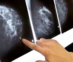
In a study of 185 women ages 40 to 50, 22% of those who had a mammogram in the year prior to diagnosis had a mastectomy, compared with 52% who did not have a mammogram in the previous year (p = 0.02).
In addition, women who had been screened in the year prior to diagnosis had smaller tumors: 17.8 mm, on average, compared with 24 mm for those who'd had mammography more than a year ago and 28.8 mm for those who never underwent screening.
They also had less likelihood of multifocal malignancy -- 12% versus 34% for those who were never screened (p = 0.003) -- and they were less likely to have high-grade disease, at 31% versus 46%, respectively (p = 0.03).
The odds of mastectomy also correlated with the size of the tumor (p = 0.04), according to Nicholas Perry, director of the London Breast Institute at Princess Grace Hospital in London.
"Our study delivers strong clinical evidence to support annual screening of women from age 40," he said.
The researchers reviewed clinical and imaging data on women who were seen at their institute between 2003 and 2009. Only 26% had mammograms prior to their diagnosis.
Of the 48 women who had ever had a mammogram, 18 had one within the year prior to diagnosis, 15 had the exam one to two years prior, and 15 had a mammogram more than two years earlier.
Perry said that the U.K. does not recommend routine breast cancer screening for women younger than 50.
"The results are striking but should not be surprising," he said, adding that he doubted they would result in a change in recommendations.
A total of 37,000 U.S. women and 7,000 British women ages 40 to 50 are diagnosed with breast cancer each year, according to Perry. Based on these rates, yearly mammograms would spare 10,000 U.S. women and 2,000 U.K. women from mastectomy annually, he said.
When asked if the annual breast screens improved overall survival, he said longer-term studies are needed.
Wednesday, November 17, 2010
The Resilient Brain

Those whose familiarity with Oliver Sacks extends only to his vivid book titles — “The Island of the Colorblind,” “An Anthropologist on Mars,” “The Man Who Mistook His Wife for a Hat” — may picture his writing as a gallery of grotesques, a parade of the exotically impaired. Sacks, a practicing neurologist, does specialize in case studies of highly unusual patients. But even as he entertains and diverts with his dramatic tales, Sacks has always been up to something else: he is gently educating us about the frailties and flaws — and the strengths and capacities — of “normal” people, those whose afflictions are of the most ordinary sort. You may never have confused your spouse for an item of outerwear, but have you ever failed to recognize the face of an acquaintance? Fumbled for a word that eluded your grasp? Read a sentence three times and still didn’t get it?
Such familiar slips, and how we handle them, are the stealth subjects of Sacks’ latest book. “The Mind’s Eye” is a collection of essays — some of which have already appeared in The New Yorker — but it has a remarkably graceful coherence of theme, tone and approach. Once again, Sacks explores our shared condition through a series of vivid characters: the woman who couldn’t talk, the man who couldn’t read, the “prosopagnosic” who couldn’t identify her own face in a photograph. (For those who wonder just how Sacks locates such people, it soon becomes clear that many of his patients find him, after recognizing themselves in his writing. They enter his care through the pages of his books, and in turn become characters in his next round of stories.)
The sufferers who write to Sacks receive a deeply empathetic response. Of one correspondent, a woman who has lost the capacity to read (but, remarkably, retains the ability to write), Sacks notes that he responded to her by telephone. “I normally would have written back,” he tells us, but in this case calling “seemed to be the thing to do.” Over time this patient, afflicted with a degenerative brain condition called posterior cortical atrophy, loses her ability to recognize objects and people, though she retains a keen sense of color and shape. When Sacks meets her in person to see how she navigates her everyday life, he dresses head to toe in red so she can keep track of him in a crowd.
Given to such un-self-consciously generous gestures, Sacks would seem to be the ideal doctor: observant but accepting, thorough but tender, training his full attention on one patient at a time. For the patient’s benefit and for ours, he illuminates every uncanny detail, brings out every excruciating irony. The woman for whom Sacks dresses in red, for example, is a virtuoso pianist, and the first sign of her malady is a sudden inability to read music. She is joined in these pages by a novelist who wakes up one morning unable to read, and an intensely sociable woman who is suddenly struck dumb. But Sacks is not primarily interested in documenting pathology, or even curing disease, which in most cases is impossible. There are no miraculous “awakenings” here.
Rather, he is most engaged by the process of compensation, how people make up for what they have lost, wresting new possibilities from their newly imposed limits. There’s the blind man who develops super-sensitive hearing, the deaf woman who catches tiny shifts in facial expression — and that pianist, who loses her ability to read music but gains new richness in her thinking about music. “She felt that her musical memory, her musical imagery, had become stronger, more tenacious, but also more flexible, so that she could hold the most complex music in her mind, then rearrange it and replay it mentally, in a way that would have been impossible before,” Sacks writes.
Sometimes these compensations are biological, he explains. The brain, plastic even into adulthood, reshapes itself to fit a new reality. In people who become blind as adults, Sacks notes, the part of the brain that once processed visual information does not atrophy, but is reallocated for another use. “The visual cortex, deprived of visual input, is still good neural real estate, available and clamoring for a new function.”
At other times, compensation takes the form of an ingenious tool. The social butterfly rendered mute by a stroke uses a lexicon, a book full of words to which she can point. (The lexicon is devised for her by a speech pathologist who is herself, Sacks notes in passing, a quadriplegic.) The novelist employs a journal-like “memory book” to teach himself how to read again. Such tools can help forge a new whole from patients’ shattered identities. As the novelist puts it, “The memory book returned a piece of myself to me.”
Sacks is most attuned to the psychological and emotional adjustments patients make to their new status; he clearly admires how they have gone on “to develop other ways of doing things, capitalizing on their strengths, finding compensations and accommodations of every sort.” In her piano playing, Sacks writes, the woman who could no longer read “not only coped with disease, but transcended it.”
So rewarding are the compensations of Sacks’ patients, in fact, that we begin to feel as if the tragedies that befell them were not tragedies at all, but — as the self-help books say — opportunities for growth. Then we arrive at the book’s penultimate essay, about Sacks’ own ocular cancer. His story is told in journal entries, dated from December 2005 to December 2009, which take on a deepening urgency as we experience along with him one event after another: the strange symptoms, the grim diagnosis, the painful treatment, the halting, incomplete recovery. Sacks’ jaunty confidence and sanguine attitude disappear, replaced by a panicked and sometimes piteous voice that is new to the reader and (if I may be so ungenerous) quite unwelcome. On Dec. 25, 2005, he writes: “Everyone says ‘Merry Christmas!’ and I reply in kind, but this is the darkest Christmas I have ever known. The New York Times today has pictures and stories of various figures who have died in 2005. Will I be among those figures in 2006?”
I found myself longing for the return of the ideal doctor of earlier chapters, and then I saw. He was right there, teaching us one more lesson: that compensation is meager consolation, that loss is painful, no matter what replaces it. Even those of us who have never lost our sight or faced a cancer diagnosis know how profoundly unsettling change can be. A move to a new job or a new neighborhood may make us, for a time, full of complaint and self-pity. It is characteristic of Sacks’ generosity to his patients that he allows only himself to be seen in this light.
Yet Sacks does eventually rally, his playful spirit intact. He notices that the blind spot, or scotoma, in his tumor-stricken right eye resembles the shape of Australia, “complete with a little bulge in the southeast corner — I thought of this as its Tasmania.” He observes that if he keeps his gaze steady for a few moments, his brain will “fill in” his blind spot with imagery borrowed from the parts of the scene he can see. The ever resilient brain, he remarks, “does not just fill in color, it fills in patterns too, and I enjoyed experimenting with my own scotoma, testing its powers and limitations.” Sacks calls this activity “scotomizing.”
Irrepressible though he may be, Sacks will not let us forget the sober lesson of his experience. He ends the essay not with a cheering paean to human resilience, but with a bleak new turn of events. A hemorrhage has further clouded his vision, leaving him with a gaping “nowhere” in his right visual field. “Time will tell whether I am able to adapt to this new visual challenge,” he writes.
Perhaps Sacks will take comfort from his novel-writing patient, who with great effort taught himself to read again. “The problems never went away,” the novelist reports, “but I became cleverer at solving them.”
Saturday, July 31, 2010
Ewing Sarcoma
Ewing sarcoma (ES) and primitive peripheral neuroectodermal tumor (PNET) were originally described in the early 1900s as distinct clinicopathologic entities. It become evident that these entities are actually part of a spectrum of neoplastic diseases known as the Ewing sarcoma family of tumors (EFT), which also includes extraosseous ES (EES), adult neuroblastoma, malignant small-cell tumor of the thoracopulmonary region (Askin's tumor), paravertebral small-cell tumor, and atypical ES.
Because of their similar histologic and immunohistochemical characteristics and shared nonrandom chromosomal translocations, these tumors are considered to be derived from a common cell of origin. Although the histogenetic origin has been debated over the years, evidence from immunohistochemical, cytogenetic, and molecular genetic studies supports a neuroectodermal origin for all EFT
The EFT can develop in almost any bone or soft tissue, but is most common in the flat and long flat bones; patients typically present with localized pain and swelling. Although overt metastatic disease is found in fewer than 25 percent at the time of diagnosis, subclinical metastatic disease is assumed to be present in nearly all patients because of the 80 to 90 percent relapse rate in patients undergoing local therapy alone. As a result, systemic chemotherapy has evolved as an important component of treatment
Advances in multidisciplinary management of EFT over the past 30 years have resulted in a marked improvement in survival and a greater likelihood of limb-sparing surgery rather than amputation
In data derived from the Surveillance, Epidemiology and End Results (SEER) program of the National Cancer Institute, five year survival rates for patients with ES rose from 36 to 56 percent during the periods 1975 to 1984 and 1985 to 1994 .
With modern multidisciplinary treatment, long-term survival can be achieved in 70 to 80 percent of patients presenting with nonmetastatic disease
Clinical Presentation:
Primary sites — ES most often arises in the long bones of the extremities (predominantly the femur, but also the tibia, fibula and humerus), and the bones of the pelvis. The spine, hands, and feet are affected considerably less often .
Within the axial skeleton, tumors arose from the pelvis, chest wall, spine/paravertebral region, or head and neck in 45, 34, 12, and 9 percent of cases, respectively. Compared to undifferentiated ES of bone, PNET and EES more often arise within the axial rather than the appendicular skeleton
Approximately 25 percent of patients have a soft tissue primary.
Signs and Symptoms :
Patients with EFT typically present with localized pain or swelling of a few weeks or months duration
Trauma, often minor, may be the initiating event that calls attention to the lesion. The pain may be mild at first, but intensifies fairly rapidly; it may be aggravated by exercise, and is often worse at night. A distinct soft tissue mass can sometimes be appreciated. When present, it is usually firmly attached to the bone and moderately to markedly tender to palpation . Swelling of the affected limb with erythema over the mass is not uncommon.
Patients with juxta-articular lesions may present with loss of joint motion, while lesions involving the ribs can be associated with direct pleural extension and large extraosseous masses .
When the spine or sacrum are involved, nerve root irritation or compression can result in back pain, radiculopathy, or symptoms of spinal cord compression (eg, weakness or loss of bowel and/or bladder control).
Constitutional symptoms or signs, such as fever, fatigue, weight loss, or anemia, are present in about 10 to 20 percent of patients at presentation . Fever is related to cytokines produced by the tumor cells, and along with other systemic symptoms, is associated with advanced disease.
Approximately 80 percent of patients present with clinically localized disease, although as noted previously, subclinical metastatic disease is presumed to be present in nearly all. Overt metastases may become evident within weeks to months in the absence of effective therapy. The significance of this lies in the frequent delay between the onset of symptoms and diagnosis, which in one report averaged over nine months .
Patients with primary pelvic tumors are significantly more likely to present with metastatic disease compared to other sites .Other factors associated with clinically evident metastatic disease at presentation include high level of lactic dehydrogenase (LDH), the presence of fever, an interval between onset of symptoms and diagnosis less than three months, and age older than 12 years
Sites of metastatic disease at diagnosis are similar to those seen with recurrent disease; lung and bone metastases predominate, in roughly equal proportions. The spine is the most frequently involved bone.
Lung metastases represent the first site of distant spread in 70 to 80 percent of cases, and are the leading cause of death for patients with EFT.
Lymph node, liver, and brain involvement are distinctly uncommon.
Radiographic Studies :
The diagnostic work-up is usually initiated with a plain radiograph of the affected area. ES involving bone typically presents as a poorly marginated destructive lesion, most often associated with a soft tissue mass. The tumors tend to be large, and in long bones are metaphyseal or diaphyseal in location.
The radiographic appearance has been described as "permeative" or "moth-eaten", indicative of a series of finely destructive lesions that become confluent over time. The cortex at the site of the lesion is often expanded, and the periosteum displaced by the underlying tumor, resulting in the clinical sign of Codman's triangle. The characteristic periosteal reaction produces layers of reactive bone, deposited in an "onion peel" appearance.

The soft tissue component of the tumor rarely shows any calcification or ossification. Sclerosis, if present, represents a secondary bone reaction rather than the primary bone formation that characterizes osteosarcoma. A pathologic fracture is present at diagnosis in 10 to 15 percent of cases.
Compared to plain radiographs, a CT scan of the primary site better delineates the extent of cortical destruction and soft tissue disease
However, definition of tumor size, local intraosseous and extraosseous extent, and the relationship of the tumor to fascial planes, vessels, nerves, and organs is best achieved by magnetic resonance imaging (MRI)
Imaging of the entire involved bone is necessary to exclude the presence of skip lesions (ie, medullary disease within the same bone, but not in direct contiguity with the primary lesion)
Prognostic Factors :
--Disease Extent: Presence or absence of metastases,patients with bones and lungs metastases fared worse than patients with bone metastases alone
--Tumor site and size : For patients presenting with localized disease, those with axial primary tumors (ie, pelvis, rib, spine, scapula, skull, clavicle, sternum) have a worse treatment outcome than those with extremity lesions
--Response to therapy: Patients with apparently localized disease have only a 10 to 20 percent likelihood of cure if treated with surgery or radiotherapy alone; this is improved dramatically when chemotherapy is added to treatment.Both the completeness of surgical resection and the response to induction therapy are important prognostic factors. Patients who are left with significant amounts of viable tumor in the resected specimen following neoadjuvant chemotherapy do worse than those with minimal or no residual tumor.
--Histology:In most but not all studies, neither the presence of neural differentiation (eg, as in PNETs) nor extraosseous origin has a significant adverse influence on outcome
--Age: contraversy about age as prognostic factor, the five-year relapse-free survival was significantly better for children younger than 10 compared to older children
--Molecular findings:deletion of the short arm of chromosome 1p, homozygous deletions of CDKN2A and p16/p14ARF, and p53 mutations have all been associated with poor response to chemotherapy and a worse prognosis
Treatment:
Systemic chemotherapy is the mainstay of therapy ,often being used before surgery .Doxorubicin ,cyclophosphamide or ifosfamide,etoposide ,vincristine and dactinomycin are active drugs .Local treatment for the primary tumor includes surgical resection ,usually with limb salvage or radiation therapy .Patients with lesions below the elbow and below the mid-calf have a 5-year survivl rate of 80% with effective treatment .Ewing sarcoma is a curable tumor even in the presesnce of obvious metastatic disease,especially in children<11 years old.
uptodate
Harrison's Internal Medicine 2008
Because of their similar histologic and immunohistochemical characteristics and shared nonrandom chromosomal translocations, these tumors are considered to be derived from a common cell of origin. Although the histogenetic origin has been debated over the years, evidence from immunohistochemical, cytogenetic, and molecular genetic studies supports a neuroectodermal origin for all EFT
The EFT can develop in almost any bone or soft tissue, but is most common in the flat and long flat bones; patients typically present with localized pain and swelling. Although overt metastatic disease is found in fewer than 25 percent at the time of diagnosis, subclinical metastatic disease is assumed to be present in nearly all patients because of the 80 to 90 percent relapse rate in patients undergoing local therapy alone. As a result, systemic chemotherapy has evolved as an important component of treatment
Advances in multidisciplinary management of EFT over the past 30 years have resulted in a marked improvement in survival and a greater likelihood of limb-sparing surgery rather than amputation
In data derived from the Surveillance, Epidemiology and End Results (SEER) program of the National Cancer Institute, five year survival rates for patients with ES rose from 36 to 56 percent during the periods 1975 to 1984 and 1985 to 1994 .
With modern multidisciplinary treatment, long-term survival can be achieved in 70 to 80 percent of patients presenting with nonmetastatic disease
Clinical Presentation:
Primary sites — ES most often arises in the long bones of the extremities (predominantly the femur, but also the tibia, fibula and humerus), and the bones of the pelvis. The spine, hands, and feet are affected considerably less often .
Within the axial skeleton, tumors arose from the pelvis, chest wall, spine/paravertebral region, or head and neck in 45, 34, 12, and 9 percent of cases, respectively. Compared to undifferentiated ES of bone, PNET and EES more often arise within the axial rather than the appendicular skeleton
Approximately 25 percent of patients have a soft tissue primary.
Signs and Symptoms :
Patients with EFT typically present with localized pain or swelling of a few weeks or months duration
Trauma, often minor, may be the initiating event that calls attention to the lesion. The pain may be mild at first, but intensifies fairly rapidly; it may be aggravated by exercise, and is often worse at night. A distinct soft tissue mass can sometimes be appreciated. When present, it is usually firmly attached to the bone and moderately to markedly tender to palpation . Swelling of the affected limb with erythema over the mass is not uncommon.
Patients with juxta-articular lesions may present with loss of joint motion, while lesions involving the ribs can be associated with direct pleural extension and large extraosseous masses .
When the spine or sacrum are involved, nerve root irritation or compression can result in back pain, radiculopathy, or symptoms of spinal cord compression (eg, weakness or loss of bowel and/or bladder control).
Constitutional symptoms or signs, such as fever, fatigue, weight loss, or anemia, are present in about 10 to 20 percent of patients at presentation . Fever is related to cytokines produced by the tumor cells, and along with other systemic symptoms, is associated with advanced disease.
Approximately 80 percent of patients present with clinically localized disease, although as noted previously, subclinical metastatic disease is presumed to be present in nearly all. Overt metastases may become evident within weeks to months in the absence of effective therapy. The significance of this lies in the frequent delay between the onset of symptoms and diagnosis, which in one report averaged over nine months .
Patients with primary pelvic tumors are significantly more likely to present with metastatic disease compared to other sites .Other factors associated with clinically evident metastatic disease at presentation include high level of lactic dehydrogenase (LDH), the presence of fever, an interval between onset of symptoms and diagnosis less than three months, and age older than 12 years
Sites of metastatic disease at diagnosis are similar to those seen with recurrent disease; lung and bone metastases predominate, in roughly equal proportions. The spine is the most frequently involved bone.
Lung metastases represent the first site of distant spread in 70 to 80 percent of cases, and are the leading cause of death for patients with EFT.
Lymph node, liver, and brain involvement are distinctly uncommon.
Radiographic Studies :
The diagnostic work-up is usually initiated with a plain radiograph of the affected area. ES involving bone typically presents as a poorly marginated destructive lesion, most often associated with a soft tissue mass. The tumors tend to be large, and in long bones are metaphyseal or diaphyseal in location.
The radiographic appearance has been described as "permeative" or "moth-eaten", indicative of a series of finely destructive lesions that become confluent over time. The cortex at the site of the lesion is often expanded, and the periosteum displaced by the underlying tumor, resulting in the clinical sign of Codman's triangle. The characteristic periosteal reaction produces layers of reactive bone, deposited in an "onion peel" appearance.

The soft tissue component of the tumor rarely shows any calcification or ossification. Sclerosis, if present, represents a secondary bone reaction rather than the primary bone formation that characterizes osteosarcoma. A pathologic fracture is present at diagnosis in 10 to 15 percent of cases.
Compared to plain radiographs, a CT scan of the primary site better delineates the extent of cortical destruction and soft tissue disease
However, definition of tumor size, local intraosseous and extraosseous extent, and the relationship of the tumor to fascial planes, vessels, nerves, and organs is best achieved by magnetic resonance imaging (MRI)
Imaging of the entire involved bone is necessary to exclude the presence of skip lesions (ie, medullary disease within the same bone, but not in direct contiguity with the primary lesion)
Prognostic Factors :
--Disease Extent: Presence or absence of metastases,patients with bones and lungs metastases fared worse than patients with bone metastases alone
--Tumor site and size : For patients presenting with localized disease, those with axial primary tumors (ie, pelvis, rib, spine, scapula, skull, clavicle, sternum) have a worse treatment outcome than those with extremity lesions
--Response to therapy: Patients with apparently localized disease have only a 10 to 20 percent likelihood of cure if treated with surgery or radiotherapy alone; this is improved dramatically when chemotherapy is added to treatment.Both the completeness of surgical resection and the response to induction therapy are important prognostic factors. Patients who are left with significant amounts of viable tumor in the resected specimen following neoadjuvant chemotherapy do worse than those with minimal or no residual tumor.
--Histology:In most but not all studies, neither the presence of neural differentiation (eg, as in PNETs) nor extraosseous origin has a significant adverse influence on outcome
--Age: contraversy about age as prognostic factor, the five-year relapse-free survival was significantly better for children younger than 10 compared to older children
--Molecular findings:deletion of the short arm of chromosome 1p, homozygous deletions of CDKN2A and p16/p14ARF, and p53 mutations have all been associated with poor response to chemotherapy and a worse prognosis
Treatment:
Systemic chemotherapy is the mainstay of therapy ,often being used before surgery .Doxorubicin ,cyclophosphamide or ifosfamide,etoposide ,vincristine and dactinomycin are active drugs .Local treatment for the primary tumor includes surgical resection ,usually with limb salvage or radiation therapy .Patients with lesions below the elbow and below the mid-calf have a 5-year survivl rate of 80% with effective treatment .Ewing sarcoma is a curable tumor even in the presesnce of obvious metastatic disease,especially in children<11 years old.
uptodate
Harrison's Internal Medicine 2008
Subscribe to:
Posts (Atom)