A 57-year-old man presented to the emergency department with painful purple toes. On the day of presentation, he had first been seen in another hospital but left against medical advice before the evaluation was complete. He had first noted a painful discoloration of his left great toe 2 weeks earlier. The pain and discoloration progressed to involve the left second and third toes (Figure 1A). During the 3 weeks preceding presentation, the patient also had intermittent blurry vision, intermittent chest pain, fatigue, anorexia, drenching night sweats, and a weight loss of 6.8 kg (15 lb). His roommate commented that thepatient had also been slightly confused.
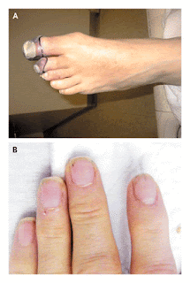
Fig1A,B
This patient's painful purple toes are suggestive of peripheral arterial ischemia. The differential diagnosis includes arterial thromboembolism resulting from a hypercoagulable state or from disseminated intravascular coagulation, perhaps related to cancer; embolus of infectious or thrombotic material from endocarditis; paradoxical embolus through an intracardiac shunt; precipitation of cryoglobulins; small-vessel vasculitis; and secondary vasculitis from an underlying connective-tissue disease, such as systemic lupus erythematosus. The possibility of cholesterol emboli should also be considered, particularly in patients who have aortic aneurysms or who have recently undergone vascular procedures. Thromboangiitis obliterans, or Buerger's disease, would be a consideration if the patient smokes. His history suggests at least two and perhaps three separate vascular events in the left foot, serially affecting the digital arteries of the great, second, and third toes. Combined with the report of blurry vision and confusion, the patient's history raises the possibility that a central vascular process is affecting the peripheral and cerebral circulatory beds. The intermittent nature of the chest pain and visual symptoms suggests the possibility of sporadic emboli. Finally, the night sweats, fevers, and weight loss point to a systemic illness, such as infection, cancer, or connective-tissue disease. Specific points to review include risk factors for subacute bacterial endocarditis, such as injection-drug use, and risk factors for hypercoaguable states, such as a family history. Further history taking and physical examination should be performed to seek evidence of hypercoagulability or a systemic illness, such as endocarditis, cancer, or vasculitis.
The patient had a history of hypertension, anxiety, chronic back pain, and gastroesophageal reflux disease. While in the Marine Corps in Vietnam, he had malaria and injuries to the hip and skull. Since serving in Vietnam he had also had post-traumatic stress disorder. Two years before presentation, a screening colonoscopy detected benign polyps. His medications included alprazolam, oxycodone with acetaminophen, and esomeprazole. The patient worked for the U.S. Postal Service and was living with a friend. He reported smoking half a pack of cigarettes daily for 30 years, weekly alcohol use, and rare but ongoing intranasal cocaine use, but no intravenous drug use. He was divorced and had not recently been sexually active. There was no family history of cancer, autoimmune diseases, diabetes, or clotting disorders.
Smoking increases the risks of several cancers, including lung and pancreatic cancers, which may in turn induce a hypercoaguable state. Intranasal cocaine use can cause vasospasm, which may exacerbate digital ischemia, and is also associated with otherbehaviors that are predisposing factors for hepatitis C, a common cause of cryoglobulinemia, and acquisition of the human immunodeficiency virus (HIV). Suspicion of limb ischemia, in addition to prompting an evaluation for physical findings indicative of such systemic disease processes, calls for a thorough examination of the peripheral pulses and may warrant consultation with a vascular surgeon.
On physical examination, the patient's temperature was 36.4°C; pulse, 104 beats per minute; blood pressure, 167/86 mm Hg; respiratory rate, 14 breaths per minute; and oxygen saturation, 96% while he was breathing ambient air. No Roth's spots were seen on funduscopy. The carotid pulses were normal, without bruits, and the chest was clear on auscultation. Cardiac examination revealed a normal S1 sound and a physiologically split S2 sound, without a murmur or rub, and the presence of an S4 gallop. The abdomen was not tender, and neither the liver nor the spleen was enlarged. There was no lymphadenopathy or thrush. The dorsalis pedis and posterior tibialis pulses in the left foot were diminished but palpable, and the foot was warm. On each of the first three toes of the left foot there was a well-demarcated, cool, tender area of nonblanching, dark-purple discoloration. Capillary refill in the unaffected toes was normal. There was no rash, edema, or livedo reticularis of the legs. Distal splinter hemorrhages were noted in several fingernails (Figure 1B). There were no Janeway's lesions or Osler's nodes. Neurologic examination revealed a deficit in the left visual field. The patient received a score of 29 points out of 30 on the Mini–Mental State Examination, having difficulty only when attempting to repeat the phrase, "no ifs, ands, or buts."
The white-cell count was 15,510 per cubic millimeter, with 80% granulocytes, 14% lymphocytes, 5% monocytes, and 1% eosinophils. The hemoglobin and platelet counts were normal, and the erythrocyte sedimentation rate was 19 mm per hour. Levels of electrolytes, blood urea nitrogen, creatinine, albumin, and globulin were normal. The level of C-reactive protein was 32.1 mg per liter (reference range, 1.0 to 3.0). The serum level of creatine kinase was 40 U per liter (reference range, 41 to 266); creatine kinase MB, 2.7 ng per milliliter (reference value, <5.0);> I, 2.22 ng per milliliter (reference value, <0.04).> normalized ratio for the prothrombin time was 1.1, the partialthromboplastin time 25.7 seconds (reference range, 23.8 to 36.6), and the fibrinogen level 264 mg per deciliter (reference range, 200 to 450). A blood smear showed hypochromia; no schistocytes were identified. An electrocardiogram showed a normal sinus rhythm with ST-segment elevations in the inferior and anteroseptal leads.
The finding of a well-demarcated, discolored area on each of the three toes of the left foot supports the possibility of a separate embolus to each of the three small digital arteries feeding the affected toes, rather than a single, more proximal thrombotic or embolic event. The lesions are consistent with localized ischemia, infarction, or thromboangiitis obliterans, but the fact that the foot is warm, with palpable pulses, rules out critical limb ischemia. Although the findings on physical examination do not rule out the possibility of a small-vessel vasculitis, the absence of livedo reticularis makes the presence of cholesterol emboli unlikely. The splinter hemorrhages are consistent with systemic emboli, but these hemorrhages are a nonspecific finding that can also be associated with nail trauma, autoimmune disease, connective-tissue disease, cancer, or endocarditis. Splinter hemorrhages are more specific for subacute bacterial endocarditis when they are present in the proximal, rather than distal, nail plate.
The elevated white-cell count suggests infection, but it is a nonspecific finding. The normal coagulation studies and absence of schistocytes on the blood smear are not consistent with a diagnosis of disseminated intravascular coagulation. The combination of the elevated level of troponin I and the ST-segment abnormalities raises the possibility of an acute coronary syndrome, although the apparent involvement of two vascular territories (right and left anterior descending coronary arteries) on electrocardiography and evidence of ischemia elsewhere make emboli to the coronary circulation seem more likely. Echocardiography might identify a cardiac source of emboli and would help to gauge the extent of myocardial injury. Brain imaging is warranted to evaluate the cause of the patient's visual field deficit and slightly altered mental status.
Magnetic resonance imaging (MRI) of the brain ), including diffusion-weighted imaging, revealed multiple small lesions in the right temporal and occipital lobes, right thalamus, both parietal lobes, left frontal lobe, and both cerebellar hemispheres; there was also a large area of enhancement in the right occipital and parietal lobes. Magnetic resonance angiography of the head and neck showed decreased blood flow in the posterior communicating artery (Fig2A,B). A transthoracic echocardiogram showed a mildly thickened mitral valve, with mild regurgitation, and structurally normal aortic, tricuspid, and pulmonary valves. The left ventricular ejection fraction was normal, and therewere no abnormalities in wall motion. The results of venous ultrasonography and computed tomographic (CT) pulmonary angiography performed at the other hospital were obtained; they showed thrombi in the great saphenous veins of both legs and a small pulmonary embolus in the lower lobe of the right lung.
 Fig-2A,B
The MRI findings are consistent with embolic disease of the brain, with the emboli mostly likely traveling through both the carotid and vertebrobasilar arteries from the left heart or proximal aorta. The disease process is not limited to the arterial circulation, as evidenced by the pulmonary embolus and venous thrombi of the legs. Further evaluation should focus on identifying a disease process that would explain both the venous and the arterial thrombosis, including evaluation for an underlying hypercoaguable state. Paradoxical embolism from a venous source, passing through an intracardiac shunt, could also explain the presence of both arterial and venous lesions. Although the transthoracic echocardiogram showed no evidence of a shunt, studies performed without the injection of agitated saline, such as this one, have a low sensitivity for atrial septal defects. Because of the high clinical suspicion for subacute bacterial endocarditis, further evaluation with transesophageal echocardiography is appropriate. Empiric antibiotic treatment should be initiated after blood cultures are obtained.
Emergency cardiac catheterization warrants consideration in any patient with ST-segment elevations, but in this case the normal ventricular wall motion on the echocardiogram, the presence of ST-segment abnormalities in two distinct vascular distributions, and the absence of ongoing chest pain all favor coronary emboli as the cause of the patient's symptoms and elevated troponin levels. Cerebral emboli are associated with a risk of hemorrhage, particularly if they are infectious, which in turn increases the risk of systemic anticoagulation, in addition to the anticoagulation that would be required with a percutaneous intervention performed at cardiac catheterization.
Anticoagulant therapy was deferred pending further evaluation. A transesophageal echocardiogram showed a 7-mm mobile echodensity on the atrial aspect of the posterior mitral-valve leaflet, with no evidence of perivalvular abscess or leaflet perforation, and mild-to-moderate mitral regurgitation (Figure 3). Treatment with ceftriaxone, vancomycin, and gentamicin was initiated for presumed infective endocarditis. However, multiple blood cultures — including cultures grown from samples obtained before antibiotic therapy was begun, cultures held for 2 weeks, and fungal blood cultures — were all negative. Serologic tests for HIV, coxiella, bartonella, treponema, and hepatitis B virus were negative. Serum hepatitis C virus RNA was undetectable.
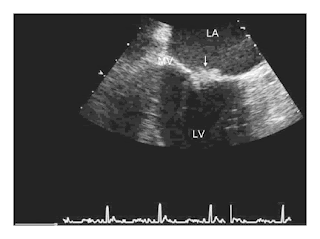 Fig3- Transesophageal Echocardiogram
A midesophageal commissural view of the mitral valve shows the lesion (arrow). LA denotes left atrium, LV left ventricle, and MV mitral valve.
The negative results of tests for infection increase my suspicion that the causes of the cardiac valvular vegetations and thromboembolic disease are noninfectious. Nonbacterial thrombotic endocarditis (commonly known as marantic endocarditis) is a potential complication of connective-tissue diseases and cancer, either of which could explain the patient's weight loss and night sweats, and may also be associated with concurrent venous and arterial thromboembolism. Further studies should include assays for the lupus anticoagulant and anticardiolipin antibodies, since the antiphospholipid-antibody syndrome could explain the valvular lesion and the venous and arterial thromboses. In the absence of localizing symptoms, imaging of the abdomen and pelvis should be considered to look for evidence of cancer.
Anticardiolipin antibodies were not detected, and a test for lupus anticoagulant was negative. CT of the abdomen revealed multiple low-attenuation lesions in the liver, a finding suggestive of metastatic disease, as well as a small low-attenuation lesion in the head of the pancreas, an enlarged gastrohepatic lymph node, and bilateral wedge-shaped renal infarcts (Figure 4). CT-guided fine-needle aspiration of a liver lesion was performed, and cytologic examination of the aspirate revealed a poorly differentiated adenocarcinoma of undetermined primary origin (Figure 5). Serum levels of alpha-fetoprotein and prostate-specific antigen were normal. The level for the beta subunit of human chorionic gonadotropin was 20 mIU per milliliter (reference range,
 1.5), and that for carcinoembryonic antigen was 4.7 ng per milliliter (reference range, 2.5). The serum level of the carbohydrate antigen 19-9 (CA 19-9) was 16,611 U per liter (reference value, <35).
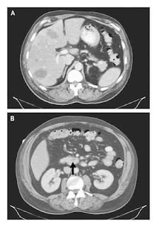
Fig4-Contrast Enhanced CT of the Abdomen
Multiple low-attenuation lesions can be seen in the liver (Panel A), and a small low-attenuation lesion is visible in the head of the pancreas (Panel B, arrow)

Figure 5.-Specimen from Fine-Needle Aspiration of a Liver Lesion.
Staining of the aspirate with hematoxylin and eosin shows sheets of poorly differentiated malignant cells (Panel A) and the mitotic figures and abundant clear cytoplasm that are consistent with adenocarcinoma (Panel B).
Although measurement of the CA 19-9 level should not be used as a screening test for pancreatic cancer in the general population because of its very low positive predictive value, in a patient with a pancreatic mass and suggestive clinical findings, such as this patient, a positive test result strongly supports a diagnosis of pancreatic carcinoma. Slight elevations in the levels of human chorionic gonadotropin and carcinoembryonic antigen are nonspecific and can be seen in pancreatic cancer, especially when it has metastasized to the liver. The identification of metastatic adenocarcinoma confirms the diagnosis of nonbacterial thrombotic endocarditis associated with cancer. Although concurrent arterial and venous thromboemboli are a rare complication of cancer, they are more common in cases of nonbacterial thrombotic endocarditis, as was true with this patient. Anticoagulant therapy with unfractionated heparin should be initiated to decrease the risk of recurrent thromboembolism.
Treatment with unfractionated heparin was initiated on hospital day 8; the regimen was subsequently changed to low-molecular-weight heparin. Over the course of the next week, progressive renal insufficiency, visual impairment, and episodes of psychosis with flashbacks to the Vietnam War developed. Plans for chemotherapy were deferred because of progressive multiorgan dysfunction and the poor prognosis, even with treatment. The patient requested transition to palliative care, and he died within weeks after his initial presentation. A postmortem examination was not performed.
Commentary
This patient's evaluation reveals how a careful history taking and a broad consideration of the possible causes of seemingly disparate events — including limb ischemia, blurred vision, chest pain, and weight loss — can lead to the unifying diagnosis of a systemic condition. The patient's history, the findings on physical examination, and the radiologic studies prompted consideration of a source of systemic emboli and the initiation of empirical therapy for infective endocarditis while an evaluation for noninfectious causes was performed. A key feature of this case was the presence of concurrent arterial and venous thromboemboli, which can be attributed to only a small number of unifying diagnoses.
Once a cardiac valvular mass was discovered, the clinicians chose to delay the use of systemic anticoagulation because of the risk of intracerebral hemorrhage. There is considerable controversy regarding the risk of hemorrhage when a patient has intracranial infective emboli. Some retrospective studies have shown a high risk of intracranial hemorrhage among patients with infective endocarditis and cerebral infarction who undergo anticoagulant treatment for cardiopulmonary bypass, whereas others have not. In this case, since the patient's blood cultures remained negative and radiologic imaging suggested disease that had metastasized to the liver, it became clear that nonbacterial thrombotic endocarditis was the likely diagnosis. Although the use of anticoagulation with heparin in the treatment of nonbacterial thrombotic endocarditis has not been studied in a randomized trial, it is thought to be beneficial, especially in cases that arise as a consequence of a malignant disease, and it does not seem to increase the risk of hemorrhage in association with cerebral emboli.
The prevalence of nonbacterial thrombotic endocarditis on autopsy ranges from 0.3% to 9.3%, depending on sample preparation and the prevalence of malignant disease in the source population. Although nonbacterial thrombotic endocarditis has been reported in neonates and children, frequently in association with congenital heart disease, it is most common in patients 40 years of age or older.In adults, it is often associated with cancer, but it has also been reported in association with systemic lupuserythematosus, burns, HIV infection, tuberculosis, uremia, radiation exposure, snakebites, and trauma from pulmonary catheters.When malignant disease is present, adenocarcinoma of the pancreas is cited as the most common primary cancer, as was most likely in this case; other cancers frequently found in patients with nonbacterial thrombotic endocarditis include lung, colon, and prostate cancers. The thrombophilia associated with malignant disease is thought to play an important role in the formation of valvular lesions; in a series of autopsy-proven cases of nonbacterial thrombotic endocarditis, disseminated intravascular coagulation was present in 71% of the cases. Although the laboratory findings in this patient were not typical of those associated with a consumptive coagulopathy, such as the presence of schistocytes, elevated clotting times, low fibrinogen levels, and low platelet levels, the pulmonary embolus and the venous thrombi in the legs were consistent with a hypercoaguable state induced by malignant disease.
The pathophysiology of nonbacterial thrombotic endocarditis is not well understood. Damage to the valvular endothelium is considered to be a critical first step in its pathogenesis, and patients with rheumatic or congenital heart disease are at elevated risk. Endothelial damage may be the result of high blood flow, direct trauma, immune-complex deposition, or complement activation, or it may be an elaboration of interleukin-1, interleukin-6, and tumor necrosis factor by tumor cells. The underlying thrombogenicsurface then acts as a nidus for platelet aggregation and fibrin deposition and leads to the formation of small verrucae, most of which are less than 3 mm in diameter.The valvular lesions of nonbacterial thrombotic endocarditis are usually present on the atrial surface of the mitral valve or on the ventricular surface of the aortic valve, at the point of valve coaptation. These lesions embolize frequently; the spleen, kidney, brain, and heart are the most frequently affected organs.
Current guidelines suggest that patients with nonbacterial thrombotic endocarditis and thromboembolism should be treated with full-dose heparin.As in the management of venous thromboembolism in patients with cancer, warfarin is less effective than heparin(unfractionated or low-molecular-weight) in the treatment of nonbacterial thrombotic endocarditis.Treatment of the underlying cause of the endocarditis is most likely to lead to a cure. Unfortunately, as in the present case, nonbacterial thrombotic endocarditis is often a sign of widely disseminated cancer and carries a poor prognosis.
There are no pathognomonic features in nonbacterial thrombotic endocarditis. Fever, cardiac murmur, leukocytosis, and elevated levels of C-reactive protein are present less frequently in patients with nonbacterial thrombotic endocarditis than in those with infective endocarditis. In this case, the final diagnosis of nonbacterial thrombotic endocarditis with underlying adenocarcinoma was established only after a thorough search for a cause of both the arterial and the venous thromboses.
@NEJM
No comments:
Post a Comment